This website is intended for healthcare professionals only.
Take a look at a selection of our recent media coverage:
11th February 2025
Speaking at Hospital Healthcare Europe’s recent Clinical Excellence in Respiratory Care event, Dr Zaheer Mangera shared insights into the evolution of the neoadjuvant lung cancer pathway, its benefits and challenges, and notable changes to the TNM standards used to classify malignant tumours.
Click here to read part one of Dr Mangera’s overview of lung cancer management and diagnosis, including insights into the latest guidance, diagnostic and treatment targets and a deep dive into lung cancer screening and incidental findings.
The neoadjuvant lung cancer pathway has triggered a paradigm shift in how we’re treating our patients. About 20-25% of patients with non-small cell lung cancer have resectable disease at the time of presentation, yet 30-55% of patients undergoing curative surgery have recurrence.
What can we do to try to improve the cure rates in these groups? Historically, there’s always been some modest success with both adjuvant and neoadjuvant approaches.
A big study, CheckMate 816, looked at patients with Stage 1B lung cancer, with a tumour size of over four centimetres without any nodal metastases, to anything up to Stage 3A.
The researchers looked at patients deemed to have resectable disease at the time of multidisciplinary team (MDT) input, with a very good performance status of zero or one. This would be the first instance of having cancer treated, and they shouldn’t have an anaplastic lymphoma kinase/epidermal growth factor receptor (EGFR) mutation.
They were offered nivolumab immunotherapy, regardless of their programmed death-ligand 1. Alongside, they had a platinum-based chemotherapy versus just chemotherapy alone. They then restaged patients then proceeded to surgery.
The primary endpoint was looking at event-free survival and pathological response to treatment. It found clear difference in the event-free survival curve between those who had nivolumab with chemotherapy versus those who had chemotherapy alone. A statistically meaningful difference was sustained well beyond the three-to-four-year period.
It’s now part of NICE guidance, albeit not part of the lung cancer guidance specifically, as it’s a separate NICE technological appraisal (TA876). It’s really made us think about how we can get these patients through a pathway and the challenges involved.
The first patient on this pathway at my hospital had excellent response to the neoadjuvant treatment, then went on to have an uncomplicated surgery and now is just on standard follow-up.
Trying to give chemotherapy before surgery makes an already complex pathway even more complex. It lengthens the pathway considerably, because those three cycles can take over two months to get through, plus the required follow-up scans.
So, although the first treatment would have been given, the time it takes for you to then close the loop at your MDT and say the whole treatment has been finished is definitely elongated.
For most patients, if you offer them surgery, they want a tumour out. Of course, there are lots of other tumour groups where you give neoadjuvant treatment and so having discussions about the process is not new to the oncologist, but it’s new to our surgeons.
There are risks of toxicities, although the trial data reports that the risk is not excessive, and patient tolerance is usually very high. It does mean we need to get an EGFR status as rapidly as possible. If you’re waiting a full four weeks for the EGFR status, before you can start treatment, it can mean considerable wait time to treatment in patients who have high-risk disease – those who are on the cusp of cure and not being cured.
We’re developing rapid EGFR pathways through our pathology labs where you can get the EGFR done more rapidly or even sometimes using the CtDNA blood tests.
And how do you standardise all these care plans and pathways across all cancer alliances and Trusts to ensure everyone is on board, everyone eligible for this treatment is getting it and to ensure we really accurately record it? It’s a real cornerstone of the National Lung Cancer Audit in how we collect our data now.
NICE guidance tells us that anyone with a PET-added lymph node, or a lymph node greater than 10 millimetres, should have an endobronchial ultrasound (EBUS). This sounds reasonable, but we know from different trials that there can be a discrepancy between PET and actual lymph node sampling itself, with false negatives and false positives.
This isn’t ideal if somebody is on the cusp between having radical surgery or needing combination therapy, for example, with neoadjuvant treatment. We know that even with PET/CT and EBUS, you can still miss some patients with nodal disease, and it only becomes evident at time of surgery when you’re removing the entire node.
Accurate staging is important. There are different ways of deciding which lymph nodes are higher risk. Sometimes it’s at ultrasonography when you recognise the risk. At the time of EBUS, you can accurately measure the size, which can be more accurate than CT, PET and PET/CT. And you may well see that the nodes are actually a conglomerate of nodes, for example.
The shape can be helpful: is it oval versus round? Are the margins clear? This is a really good one because very often when you can’t find the lymph node or you can’t really see the margins, it can sometimes be reassuring compared to where you see distinct margins.
Is it homogenous, does it echo-signal consistently throughout or have subtle changes between one area and the next? Is the central hilum structure present or is it absent? Is there any evidence of necrosis?
In future, we’re largely trying to transition to any lymph node over 10 millimetres on CT being biopsied, as well as any lymph node with fluorodeoxyglucose avidity and any lymph node with high-risk features. The only way you can do that is by doing an EBUS.
The TNM 8th edition is what we’re using at the moment and the TNM 9th edition, which is due to go live imminently. The changes are reasonably subtle and there’s no significant ground shift in how we stage lung cancers.
First of all, N2 lymph nodes are going to be split into N2a and N2b. Currently it’s just N1, N2, N3 and we’re now going to have a sub-category for lymph node station N2, with N2a being single station involvement and N2b being multiple station involvement.
It doesn’t really comment too much on size here, so it still does need a little bit of working out in MDT with what you’re going to do with different lymph node groups and how it may or may not change your management and approach to the patient.
Then you’ve got your M stages. M1a and M1b are largely unchanged but M1c is split into M1c1 and M1c2.
M1c1 is multiple extrathoracic metastases in a single organ system, so multiple liver or brain metastases, rather than single metastases, and M1c2 is extrathoracic metastases in multiple organ systems.
This will subtly change the staging and may make a difference to what your approach might be. Having metastases doesn’t mean you don’t have radically treatable disease, and our oncology teams have specific MDTs for patients with metastatic disease and stereotactic ablative radiotherapy MDTs.
More and more of these metastatic diseases can be treated and that’s why it’s important to differentiate between M1b, M1c1 and M1c2 because some of these patients, particularly with M1b may well still have radically treatable disease if it’s just a single metastasis in a single organ.
Dr Zaheer Mangera is a respiratory consultant and the lung cancer lead at North Middlesex University Hospital, now part of the Royal Free London NHS Foundation Trust in London, UK.
View the agenda and register for our next Clinical Excellence in Respiratory Care event now.
30th January 2025
An ultrasensitive blood test which detects even low levels of circulating tumour DNA (ctDNA) could help to improve disease stratification in early-stage lung adenocarcinoma (LUAD), a UK study suggests.
It was known that ctDNA detection could predict clinical risk in early-stage tumours, the UK research team wrote in the journal Nature Medicine. However, it was challenging to detect preoperative ctDNA in early-stage LUADs due to low levels of ctDNA in plasma – frequently below 100 ppm.
In the Cancer Research UK-funded study, scientists from University College London (UCL), the Francis Crick Institute, University College London Hospitals NHS Foundation Trust (UCLH), and the biotechnology firm Personalis, collaborated to test the company’s NeXT Personal platform.
Described as an ultrasensitive, tumour-informed liquid-biopsy platform, the researchers said NeXT Personal had been analytically validated for ultrasensitive ctDNA detection at 1-3 ppm of ctDNA with 99.9% specificity.
They used the platform to analyse preoperative ctDNA in 171 adults with early-stage lung cancer from Cancer Research UK’s TRACERx study.
It detected ctDNA preoperatively in 81% of the patients with LUAD, including 57% of those with pathological tumour-node-metastasis (pTNM) stage 1 disease.
Analysis showed people with a low level of ctDNA before surgery were less likely to relapse and also had improved overall survival rates compared with people with a high level of ctDNA.
The researchers were also able to show that patients with <80 ppm preoperative ctDNA levels had reduced overall survival compared with ctDNA-negative patients with LUAD.
‘Although prospective studies are needed to confirm the clinical utility of the assay, these data show that our approach has the potential to improve disease stratification in early-stage LUADs,’ the study authors concluded.
The data from TRACERx was analysed retrospectively, the researchers acknowledged, with future data from prospective cohorts needed to evaluate the clinical utility of the assay.
‘Although NeXT Personal is already in use as a clinical diagnostic test, it, like other tumour-informed ctDNA detection assays, is of higher complexity and requires a longer turnaround period to develop the panel and obtain a clinically actionable result, compared with non-tumour-informed approached,’ they wrote.
Study first author Dr James Black, a postdoctoral clinical fellow at the Francis Crick Institute and the Cancer Research UK Lung Cancer Centre of Excellence at UCL, said the study had shown the presence or absence of tumour DNA in the blood was strongly predictive of prognosis.
‘ctDNA testing, especially using ultrasensitive platforms, could help clinicians make more informed decisions about treatment and give patients a more accurate idea of how their disease might progress,’ he said, adding that ‘more research to validate these tests will help to get them on the agenda for regular clinical use.’
Study senior author Professor Charles Swanton, who holds positions at the UCL Cancer Institute, the Francis Crick Institute and UCLH and is chief investigator of the TRACERx study, noted that lung cancer is one of the most common types of cancer in the UK and has a high relapse rate.
‘It’s vital to understand who would benefit from more aggressive treatment, especially for patients with stage 1 disease who are often diagnosed during CT screening for those at a higher risk,’ he said.
‘Using sensitive ctDNA tests is one way to do this, which we hope will maximise clinical benefit and minimise unnecessary treatment for individual patients.’
Speaking at Hospital Healthcare Europe’s recent Clinical Excellence in Respiratory Care event, Dr Zaheer Mangera,the lung cancer lead at North Middlesex University Hospital NHS Trust in London, said ctDNA tests were among the innovations to watch in lung cancer care, with the Trust involved in a ctDNA pilot in recent months.
17th January 2025
Speaking at Hospital Healthcare Europe’s recent Clinical Excellence in Respiratory Care event, Dr Zaheer Mangera shared insights into the lung cancer pathway and its management and diagnosis in accordance with NICE and other guidance, paying particular attention to the benefits and challenges of early diagnosis and lung cancer screening.
Cancer waiting times are never far from the headlines, and recent figures on these targets for all cancers show mixed success, with the target for the faster diagnosis standard being met (77.4% vs 75%) but the 31-day decision to treat and 62-day referral to treat standards falling short of their respective targets (91.0% vs 96% and 69.4% vs 85%).
When it comes to lung cancer, more than 5,000 cases have been identified through the NHS Targeted Lung Health Check Programme since its launch in 2019. Some 76% of these were found at the earliest stages of one and two, offering those patients the best chance of survival.
In fact, NHS data shows a 7.4% improvement in lung cancer early diagnosis rates from April 2023 to March 2024 compared to March 2019 to February 2020. However, the overall picture is varied and there’s still work to be done to ensure targets are met, screening successes continue and patients receive the best treatment as early as possible.
Respiratory consultant Dr ZaheerMangera is the lung cancer lead at North Middlesex University Hospital – now part of the Royal Free London NHS Foundation Trust – in London, UK. Optimising and delivering a successful lung cancer pathway is one of his main focuses, and this requires a careful balance of speed, accuracy and resource.
There hasn’t been much dramatic change to NICE guidance – since 2019, it’s been more subtle. Some of the big changes have been around early diagnosis, specifically lung cancer screening, which I’ll come onto shortly.
The way I look at NG122 is that it provides a basic framework, setting down the minimum criteria for delivering a lung cancer pathway. Lots of the guidance relates to order, process and speed. It doesn’t really provide a manual of how you should treat lung cancer, but it does touch on some important aspects.
Faster diagnosis consumes us as lung cancer physicians – everything is about speed, getting patients discharged off the pathway as quickly as possible, and trying to get those who do have lung cancer through it so they can get treatment at lightning speed.
The document Millimetres Matter, published by the United Kingdom Lung Cancer Coalition in 2018, is an important first stepping stone in terms of speeding up the lung cancer pathway. It recognises that when looking at the T-stage of a tumour, the stage of a patient can change just by a millimetre. And those changes can happen within the course of a 62-day lung cancer pathway.
We want everyone to be sprinting towards that finish line, whether it’s discharging from the pathway or making a lung cancer diagnosis. But we know from numerous lung cancer audits year-on-year that there’s huge variety of performance. Some Trusts are doing an excellent job getting patients through the pathway rapidly, others not so good.
But this speed is important as this report highlights a 16% mortality increase if the time from diagnosis to surgery goes beyond 40 days. So, it’s important we challenge ourselves to 62 days, knowing significant numbers of patients’ cancers will progress within the lifetime of that pathway.
Patient experience is really important. Sometimes my patients say everything’s going too quickly for them, but most want to get their diagnosis the day before yesterday.
Many would have been to their GP or emergency department several times before even arriving on the pathway. It’s all about trying to consistently drive standards up and recognise what specific standards we should be focusing on to even out performance across the different Trusts.
With the lung cancer pathway, Trusts aren’t actually competing with each other, but we want them to be within a hair’s breadth of each-other – like in the Paris Olympics 100m final – because we don’t want to see an ongoing postcode lottery as to whether patients survive the pathway and get access to the best treatments within a rapid timeframe. This all coincides with the faster diagnosis standard.
A lot of my time is spent looking at how many patients are breaching the 62-day pathway and how many are inside it, but the faster diagnosis standard is important. It’s quite a simple principle: within 28 days of the referral being sent by primary care, the patient needs to know whether they have a cancer or not. They don’t necessarily need to know the treatment plan or next steps, but they need to be told that much.
At my Trust – a medium-sized district general hospital in London – the last audit showed that for every 100 GP referrals, there were three cancers found, meaning there are 97 patients sitting there worrying they may have lung cancer. So, the benefits are huge if we can achieve this 28-day target of telling patients.
If we look at the National Optimal Lung Cancer Pathway (NOLCP), we’re expecting Trusts to ensure that the patient has had an X-ray, ideally, before they’ve even seen us. We’re also looking at the CT being done within 72 hours of that referral.
We’re failing at this in my Trust quite abysmally at the moment, which is really disappointing. It’s even more disappointing that we were achieving this pre-Covid and so post-Covid there has clearly been a catastrophic collapse in how we arrange and deliver our radiology. There are lots of different factors behind that, but we certainly haven’t been able to achieve this CT target for quite some time.
But the way we mitigate that is by trying to fast-track high-risk patients, particularly those with an X-ray abnormal for lung cancer specifically. Something being developed in a number of Trusts, which I think is live in Manchester, is artificial intelligence (AI) reporting of X-rays. If there is an abnormality, it can get flagged for reporting by a radiographer or radiologist earlier on. This is one area where we may be able to pick up these high-risk cancers much more quickly.
Ideally, we want patients to meet a lung cancer specialist – or a clinical nurse specialist in Trusts that have them – within the first six days. By day 14, we want the whole panel of tests done: PET/CT, if relevant, spirometry and more advanced lung function tests like gas transfer.
Then by day 21, we want that full multidisciplinary team (MDT) discussion where we make a treatment plan, so by day 28 we’re giving the patient the all-clear or telling them the diagnosis. That gives plenty of time to get treatment started by day 49 – the maximum length of the NOLCP.
The paradigm shift is that rather than giving ourselves 62 days, we’re trying to get that treatment within 49 days, which is what we’re increasingly being audited against. Although the National Lung Cancer Audit is still publishing the 62-day results, but it’s the 49 days that’s quite important now.
More hospitals have endobronchial ultrasound (EBUS) as part of their suite of investigations compared to a decade ago, so there’s less referral into other centres. But time to EBUS can still be problematic and patients in some areas, particularly for general anaesthetic EBUS procedures.
We’ve also seen a whole range of barriers in my own Trust around PET/CT scans. We’ve been waiting three to four weeks for a PET/CT and more recently have got them done within three to four days as more scanners come online. The issues are the physical infrastructure required to get a PET/CT scanner in, radiology reporting limitations and the fact that there won’t be this scanner in every hospital – there is going to be a hub and spoke model.
Perhaps most pertinent barrier is getting access to molecular markers for treatment plans, epidermal growth factor receptor (EGFR) status and immunohistochemistry. In my practice, it can be a full four weeks from the day I take the biopsy before we’ve got access to the full molecular markers.
Another innovation is access to circulating tumour DNA (ctDNA) blood tests. It’s been online for over a year, and my Trust has been part of a pilot for the last six months. A simple blood test is sent to a specialist lab and you get a very detailed report within two weeks. It’s particularly good if you’ve got somebody with stage three cancers or above where there’s metastases and some tumour DNA has spilt into the blood. We’re getting quite a few false negatives where the patient may well have cancer, but there’s just not enough tumour DNA present for any meaningful results to be gained from the blood test. But this can speed things up, and you can use it alongside your biopsy results to determine appropriate treatments.
The first thing to say about early diagnosis is that within the NHS, there is almost a pseudo screening programme, given how many patients are receiving all kinds of CT scans that incidentally pick up early lung cancers.
These are all very fruitful pathways for us lung cancer physicians because we tend to pick up asymptomatic lung cancer from, say, the chance CT colonogram that may have included a CT chest as part of its protocol. There are hundreds of CT coronary angiograms being done every month at our local tertiary cardiology centre and we’ll see a small percentage of them, but it actually ends up being quite a large number with reported nodules.
For those of you working alongside rapid diagnostic centres where, typically, patients will present with symptoms of weight loss, but no clear pathway for them to be referred to, they’ll be churning out quite a lot of CT chest-abdomen-pelvis. There’s direct access to CT pancreas in many areas for GPs, which can sometimes include a CT chest – the list goes on. We’re seeing lots of imaging, and we’re finding lots of incidental findings, and it’s quite a rich resource.
The message is that all lung MDTs – whether diagnostic or treatment – are absolutely being flooded with incidental findings but as some of these are picking up early lung cancers, it’s very difficult for us to say we want that Pandora’s box to be closed and we want this imaging to be a bit more thoughtful and a bit more targeted. This is going to be an area that becomes increasingly important for us to navigate.
Lung cancer screening has captured the imagination of the UK, particularly of England, and we’re seeing more lung cancer pathways. Some are hospital-based, some are in community hospitals, and some are roaming around with patients sometimes being invited to screening on lorries in supermarket car parks.
The lung cancer screening story started back in the 1970s, but real major changes to practice were first initiated by a study initiated in the 2000s and reported on around 2010/11. Here, America’s National Lung Screening Trial found you could secure a 20% reduction in lung cancer mortality if you started screening high-risk patients, typically smokers between age 50 and 70. Subsequent studies, like the European Nelson Study, found a mortality reduction of up to 26%.
It answered the question, beyond any reasonable doubt, that if you want to try to improve cancer mortality, earlier diagnosis is the strategy, and you need an appropriate tool to do that. A non-contrast, low-dose CT is a very effective way of ruling in or ruling out lung cancers.
One interesting outcome is a differential in the mortality gain between men and women. In the Nelson Study, for example, you can get up to 33% improvement in mortality in females compared to males where it’s around 25%, and it’s listed as a risk reduction.
This doesn’t necessarily change our approach, but it tells us a bit about the biology of cancer in women, and how they may well have more treatment options and better response to treatments. But remember, this population are smokers or ex-smokers, so it doesn’t answer the whole question about the difference between men and women but it’s an interesting observation.
In the UK, you have to be between the age of 55 and 75, you need to be registered with a GP, and you need to have a smoking history. Different risk assessment tools or prediction models are used – whether it’s LLPv2 or a PLCO – and, depending on the risk score, you can be invited for a CT scan screening.
There are lots of challenges around targeted lung health checks and the first is how many patients are agreeing to have a scan. CT scans aren’t usually as problematic as other types of screening – such as bowel screening – and may feel less invasive for some patients, but they still require the patient to engage, taking time off work or travelling quite some distance in some cases.
Nationally, the most recent update reveals around 42% of patients have a CT scan when invited. Some areas, like North East London, for example, have an uptick of around 80%.
It all depends what strategies are in place to try to improve uptake and how you can engage with your community and engage with people who may be at risk, including people without English as their first language and other groups that are more difficult to reach.
The number of nodules that our colleagues are finding in these targeted health checks is monumental. AI reporting and having a clear protocol for these does help, and a lot of these incidental findings can be dismissed without ever coming directly into an NHS lung cancer pathway.
In my experience, we’re only seeing those who are genuinely higher risk or are borderline. We’re not seeing too many patients who just need a standard lung nodule follow-up.
It’s a challenge in terms of aligning clinical systems and ensuring everything works, given most hospitals don’t have integrated systems. You’ll have a targeted lung health check serving a number of different hospitals that all have different systems that don’t talk to each other.
Where I work, we’re still receiving referrals by email, which of course is problematic if an email isn’t read or actioned in the usual way.
There are questions over whether we should be scanning younger or older patients and that will always be a big debate – have we got the age groups correct?
And this is all being done in the context of an under-resourced lung cancer service. Looking at the national picture, our mortality rates are still well behind most of Europe. A lot of this is to do with the actual resources and numbers of PET/CT scanners, chest physicians and oncologists.
We can identify other life-threatening findings, like an aortic aneurysm that’s about to rupture, for example. Patients will get spirometry during the testing phases as well, so you can diagnose COPD. The CT findings themselves can offer important lung findings, such as undiagnosed pulmonary fibrosis or other interstitial lung diseases – bronchiectasis, for example – and cardiac conditions such as the degree of calcification of the coronary arteries.
It’s also an opportunity to offer a tobacco dependency service, because many of these patients will be current smokers, and so it’s a good way of offering a treatment for their tobacco dependency.
Click here to read part two of Dr Mangera’s overview of lung cancer management and diagnosis, which focuses on the evolution of the neoadjuvant lung cancer pathway and notable changes to the TNM standards used to classify malignant tumours.
View the agenda and register for our next Clinical Excellence in Respiratory Care event now.
17th October 2024
Hospital Healthcare Europe (HHE) is delighted to welcome respiratory consultant Dr Zaheer Mangera as a speaker on the diagnosis and management of lung cancer at the upcoming Clinical Excellence in Respiratory Care event on 25 November 2024.
Dr Mangera, who is the lung cancer lead at North Middlesex University Hospital NHS Trust, will touch on guidelines from the National Institute for Health and Care Excellence as he considers the importance of early diagnosis and indications for chest radiotherapy, helping patients to understand what their risk factors are and what interventions are available to them, plus the effectiveness of diagnostic and staging investigations.
This session complements his contribution to the previous Clinical Excellence in Respiratory Care event in May 2024 in which he joined a panel discussion on the use of diagnostic imaging for respiratory conditions, with a particular focus on lung cancer.
Dr Mangera will also join colleagues from Sheffield Teaching Hospitals NHS Foundation Trust and Royal Brompton Hospital for a panel discussion on the management of pulmonary hypertension as a multidisciplinary team.
Find out more about the upcoming Clinical Excellence in Respiratory Care event and register for free to hear the latest from Dr Mangera on lung cancer and more.
This latest event in HHE’s Clinical Excellence series brings together renowned experts from recognised Centres of Excellence and other key institutions to share best practice and explore the latest advances in respiratory care including diagnosing lung cancer, personalised approaches to asthma and COPD treatment and managing pulmonary hypertension and sleep disordered breathing.
Providing the opportunity to gain CPD hours, the day-long event will also focus on how to best use multidisciplinary teams (MDT) and improve patient care in this area. The agenda has been created by HHE with guidance from industry experts to offer respiratory physicians and members of the wider MDT a comprehensive overview of this broad clinical area.
To coincide with this event and others in the Clinical Excellence series, a whole host of additional content and interviews with prominent clinicians from Centres of Excellence and beyond have been published on the HHE website – look out for the orange Clinical Excellence tag in the Cardiovascular and Respiratory zones.
This includes a summary of a fascinating session from cardio-oncologist Dr Rebecca Dobson on the need and demand for cardio-oncology services, how they’ve developed over time and the current state of play in this evolving field.
The Clinical Excellence schedule for 2025 will be announced soon, including new clinical areas, so watch this space.
5th March 2024
Population-level immunogenetic variation is a factor underlying lung cancer risk in current and former smokers, a large study has found.
Researchers used genetic and clinical data from two large-scale population cohorts – UK Biobank and FinnGen – together with multi-modal genomic analyses of non-malignant and lung tumour samples to evaluate the effect of human leukocyte antigen (HLA) heterozygosity on lung cancer risk.
The analysis found heterozygosity at the HLA class II (HLA-II) loci was associated with reduced risk of lung cancer over more than a decade of follow-up, study authors wrote in the journal Science
Of note, HLA-II heterozygosity was associated with reduced risk of lung cancer in both current and former smokers, but not never-smokers.
This suggested that smoking-derived antigens might augment the immune response to early neoplastic disease, they wrote.
In contrast, HLA-II homozygosity conferred substantial lifetime risk of disease (13.9% for current smokers in the UK Biobank cohort) and was independent of known clinical and genetic risk factors.
Co-senior author Dr Diego Chowell, assistant professor of oncological sciences, and immunology and immunotherapy at the Icahn School of Medicine at Mount Sinai in New York, which led the study in collaboration with the University of Helsinki and Massachusetts General Hospital, said the findings challenged conventional thinking by demonstrating that immune genetics, specifically HLA-II heterozygosity, played a significant role in lung cancer risk, especially among smokers.
Dr Chowell said: ‘Further, when we added polygenic risk scores – which is a measure of genetic predisposition based on multiple genes – to the analysis, it increased the lifetime risk of lung cancer, specifically in smokers who have identical versions of the HLA-II genes.’
HLAs are cell surface molecules that recognise antigenic peptides and present them to T-cells to initiate an immune response.
In lung cancer, genetic variation in the HLA locus was known to be linked to tumour evolution and treatment outcomes, but it was unclear whether HLA polymorphisms reduced lung cancer risk, the researchers explained.
This study was designed to test the heterozygote advantage hypothesis, which is a foundational principle of the evolution of the HLA system and of HLA-mediated protection against disease.
‘According to this hypothesis, individuals heterozygous at HLA are afforded greater protection against disease because they present more antigens for T-cell recognition through their two different HLA allomorphs than do homozygous individuals and consequently clear infected or neoplastic cells more efficiently,’ the researchers wrote.
The data underscored the role of immunosurveillance in protecting against lung cancer, the researchers said.
‘The association of HLA-II heterozygosity with reduced risk of lung cancer implies that genetic variation in immune-surveillance is a feature of cancer susceptibility, together with environmental exposures, hereditary risk and DNA replication errors,’ they said.
The findings broadened the understanding of the role the host immune system played in cancer risk and might motivate the incorporation of immunogenetics into lung cancer screening programmes, the study authors concluded.
‘A greater understanding of immunogenetic determinants of cancer risk, including genetic variation in HLA and other immune genes and pathways commonly associated with autoimmune and infectious diseases, may foster the development of improved strategies for cancer prevention,’ they wrote.
The findings suggested that current or former smokers homozygous at HLA-II could be considered at an earlier age for low-dose computed tomographic (LDCT) screening, which might reduce lung cancer mortality.
Whether the combination of genotype-driven risk assessment and LDCT reduced lung cancer mortality compared with either method alone should be comprehensively investigated in a future prospective clinical trial, they said.
4th January 2024
With the results of the lung cancer screening SUMMIT study expected imminently, Helen Gilbert caught up with consultant respiratory physician Dr Neal Navani to discuss this research, promising new innovations in lung cancer diagnostics and what they might mean for the future of lung cancer care.
As Cancer Research UK’s Lung Cancer Centre of Excellence, University College London and University College London Hospital (UCLH) have been at the forefront of lung cancer innovations, pioneering diagnostic modalities such as endobronchial ultrasound.
This diagnostic focus is particularly pertinent as lung cancer is Europe’s biggest cancer killer, with 380,000 deaths across the continent in 2020 – a fifth of all cancer deaths.
In England, more than 60% of lung cancer patients are diagnosed at either stage three or four, and this late diagnosis is a frustration for Dr Neal Navani, lead consultant respiratory physician for lung cancer services at UCLH, as he says cure rates can be as high as 80-90% for patients whose small, early-stage lung cancer is detected.
Dr Navani, who is also the clinical lead of the UK National Lung Cancer Audit and clinical director for the Centre for Cancer Outcomes at the North Central London Cancer Alliance, has long been involved in pioneering research at UCLH to improve early detection and diagnosis.
And recent projects suggest there are further innovations on the horizon that have the potential to improve patient outcomes.
In May 2023, the largest lung cancer screening study of its kind in the UK drew to a close.
The four-and-a-half-year SUMMIT study was a collaboration between researchers from UCLH, University College London (UCL), the National Institute for Health Research, UCLH Biomedical Research Centre and GRAIL – a US healthcare company focused on the early detection of cancer.
Their aim was to identify lung cancer early among at-risk Londoners and support the development of a new blood test for the early detection of lung and multiple cancer types.
More than 13,000 people aged 55-77 from north and east London who had a significant smoking history were offered a blood test and a low-dose CT scan of their lungs. They were followed up at three months or immediately if a cause for concern was identified.
Dr Navani describes the research – results of which are expected imminently – as ‘a really fantastic, rich data set on which we can look to answer a lot of questions about detecting cancer early’.
He is particularly interested in developing a model that incorporates PET-CT scans to predict malignancy in screen-detected lung nodules. Often these appear like freckles on the lung, which may or may not be cancerous.
The challenge, he says, is working out whether they are malignant or benign, and currently this is done using a risk calculator developed in 2005.
It involves an injection of radioactive sugar before a PET-CT scan to see whether the nodule – or anything else for that matter – takes up the sugar. This then correlates with the risk of malignancy.
However, Dr Navani describes the current tool, which was developed in 2005, as out of date and prone to underestimating the risk of cancer in lung nodules.
Data from the SUMMIT trial are set to be used to develop and test a new risk calculator that takes into account more than 10 factors including family history, smoking and the size and appearance of nodules. It aims to accurately predict the chance of a nodule being cancerous.
‘We’re able to see whether sugar is taken up by that nodule in the lung – the idea being that small cancers use up more sugar than nodules that are not due to cancer,’ Dr Navani says.
‘Data for that work are being collected and developed. We’re pulling together data through other trials doing a similar thing and hopefully we’ll be able to clarify the role of PET-CT scanning for nodules in the next two years.’
The risk calculator will be compared against the existing model as well as others that do not include PET-CT scanning.
If found to be more accurate, the potential benefits are numerous and may include fewer patient investigations at lower cost, earlier treatment and reduced anxiety for those called in, Dr Navani explains.
UCL researchers are also using blood samples from the SUMMIT study to evaluate a blood test that can diagnose tumours earlier and detect 50 types of cancer, including lung cancer, with high accuracy.
Developed by GRAIL and an international team of researchers co-led by UCL, the test looks for tell-tale chemical changes to bits of genetic code – cell-free DNA – that leak from tumours into the bloodstream.
It was developed using artificial intelligence (AI) after researchers fed data on methylation patterns from the blood samples of thousands of cancer patients into a machine learning algorithm. It is said to identify many types of cancer, including bowel, ovarian and pancreatic, and can diagnose in which tissue the cancer originated with 96% accuracy.
But the potential of technology in bolstering cancer diagnosis doesn’t stop at AI. Another promising area of innovation is robotics.
Dr Navani is intrigued by the potential of this kind of diagnostic ability, and he is aware of robotic techniques that will be ‘the subject of research over the next year or two’.
He says: ‘We need to understand the cost effectiveness of robotic diagnosis of lung nodules. It’s potentially exciting.’
Earlier this year NHS clinicians at the Royal Brompton and St Bartholomew’s Hospital in London began a clinical study trialling a robotic-assisted bronchoscopy system.
Each hospital site is aiming to recruit around 50 patients with small lung nodules located in areas that are challenging to reach via traditional bronchoscopy.
The system combines software, robotic assistance and a flexible catheter with a camera to create a 3D roadmap of the lungs – much like a car’s sat-nav.
Doctors are directed to deep and hard-to-reach areas in each of the 18 segments of the lung, with the aim of removing tissue samples for biopsy with greater precision and accuracy.
The benefits of diagnosing a lung nodule accurately with a tiny camera could ‘open up a world of possibilities in terms of drug delivery, or ablation [to destroy cancerous nodules] in a controlled and accurate way,’ says Dr Navani. ‘I think in the next five to 10 years we’re going to see novel diagnosis and treatment options for our patients with early-stage lung cancer in particular.’
Another key development Dr Navani anticipates is the continued and increasing importance of collaboration, particularly when it comes to technology.
Endobronchial ultrasound (EBUS), one of the biggest innovations in respiratory medicine over the last 15 years, evolved from endoscopic ultrasound used in other clinical areas.
EBUS was trialled in the early 2000s by the UCLH research team, of which Dr Navani was a leading player, and uses a bronchoscope with a light, camera and integral ultrasound scanner to produce a detailed image inside the chest.
It enables doctors to take targeted needle biopsies of any enlarged lymph nodes and suspicious lesions while avoiding areas such as blood vessels.
Prior to this, patients at-risk of lung conditions required incisions to the chest under general anaesthetic, resulting in hospital stays and the possibility of complications or even death.
The arrival of EBUS in clinical practice in 2007 meant the diagnostic procedure could be performed on outpatients in under 30 minutes, with patients able to leave just one or two hours later.
‘It’s a very safe technique [and] in the last 10-15 years it’s really become a mainstay of diagnosis in respiratory medicine,’ Dr Navani acknowledges. ‘It started off very slowly but now in the UK there are 140 centres that are doing this technique and it’s been adopted globally for diagnosing lung conditions.’
Dr Navani believes the adaption of tools and techniques used in other clinical fields will continue to play a pivotal role in the advancement of lung cancer diagnostics and treatment. He points out, for example, that tumour ablation, which is used to treat lung and liver cancer, is now happening at a research stage for pancreatic cancer.
And this collaboration doesn’t just extend across clinical specialities. Imaging and information providers, including the likes of Fujifilm, also serve a vital purpose by providing increasingly innovative imaging solutions.
In June 2023, NHS England announced the national rollout of a targeted lung cancer screening programme to help detect cancer sooner and speed up diagnosis.
The rollout followed a successful pilot phase in which lung cancer scanning trucks carrying out on-the-spot chest scans operated from convenient locations such as football stadiums, supermarket car parks and town centres.
In September, NHS England announced that more than one million people had been invited for a lung cancer check via the scheme and almost 2,400 cancers had been caught – an impressive 76% of which were diagnosed at stage one or two.
‘That’s going to hopefully need innovative imaging solutions, particularly low-dose scanners, and I think we need to work with industry in terms of the use of artificial intelligence to help with the reporting of those scans,’ Dr Navani says.
Innovative diagnostic imaging techniques are certainly in development, and Dr Navani sees huge potential in new technologies for treating patients, too.
‘In terms of delivering novel therapies, in the future there may be a role for delivering drugs directly into the lungs, the pleural space or endobronchially, lymph nodes, or primary lung lesions,’ he says.
Dr Navani describes working in a hospital that is attached to a world-class university as ‘fantastic’ because it grants access to ‘extraordinary expertise’ spanning science, sociology, data science, computer science and engineering.
‘The research into lung cancer at UCL is really incredibly broad and, dare I say it, world leading, right the way through the basic science, biology and understanding how cancer develops and spreads and changes over time… to understanding the societal impact, equality and equity of care,’ he says.
According to Dr Navani, there appears to be a big difference in the outcomes of lung cancer patients based on socio-economic status.
‘We’ve really tried to address this in the National Cancer Audit, but it remains a significant challenge,’ he says. “A lot of this comes down to local resources… access to healthcare, equality and subsequent diagnosis and treatment in a timely fashion.’
Another major unmet need, Dr Navani says, is the 15% of patients with lung cancer who have never smoked and it’s here that ‘urgent research is needed’.
‘Given the high burden of lung cancer care, that’s a significant number of people – if you consider [non-smoking-related lung cancer] as a cancer in its own right it would be the seventh most common cause of cancer death,’ he says.
‘We’re really starting to get to grips with lung cancer in smokers but we are still at the early stages of understanding why people who’ve never smoked develop lung cancer. It would be important to predict who these people might be so that we can identify them at an earlier stage so hopefully their outcome will be better.’
The most pressing issue facing the NHS is limited resources, according to Dr Navani.
‘We simply don’t have enough scanners, radiologists, or space to do bronchoscopies,’ he states. ‘We’ve talked a lot about innovation but actually the most important thing that can be done to improve lung cancer care is for each hospital and primary care setting to have the appropriate resources to deliver what we know is already appropriate care, to drive out inequalities and drive everybody up to the best possible standards.’
While the future of funding for lung cancer care in the UK remains in flux, one thing is for certain: the research, expertise and drive to support the early diagnosis of patients remains, and Dr Navani’s commitment to supporting patients through innovative routes is stronger than ever.
19th December 2023
The UK’s devolved nations could detect greater numbers of lung cancer cases with improved screening programmes, new analyses by Cancer Research UK have found as the charity calls for governments to ’urgently implement’ such programmes.
Lung cancer is the leading cause of cancer death in England, Scotland, Wales and Northern Ireland and smokers and former smokers aged 55-74 are at greatest risk.
In November 2022, the National Screening Committee (NSC) recommended that everyone in this group be screened but, to date, only England has initiated a screening programme on this advice.
Almost 900,000 people were invited for checks during the pilot stages in England. Of the 200,000 scans carried out, more than 2,000 people were detected as having lung cancer, and 76% of those were identified at an earlier stage (stage 1 or 2) compared to 29% identified outside of the pilot programme in 2019.
Cancer Research UK’s latest analyses suggests that equivalent lung cancer screening programmes in the devolved nations could diagnose around 4,000 more lung cancer patients in Scotland at an early stage over the next decade, as well as 2,400 more people in Wales and 1,400 in Northern Ireland.
If just 50% of eligible people took part in screening, earlier lung cancer diagnosis could save 2,300 lives in Scotland, 1,000 lives in Wales and 600 lives in Northern Ireland over the next 10 years, Cancer Research UK said.
Professor David Weller, professor of general practice at the University of Edinburgh, believes a nationwide screening programme ’has the potential to be a game changer when it comes to reducing the burden of lung cancer in Scotland’.
He said: ’For too long, lung cancer has been perceived as a disease you can’t do anything about, but we know that diagnosis at an early stage really can make a difference.
’Major trials of targeted lung cancer screening show significant reductions in mortality from lung cancer. Pilot studies in the UK and internationally consistently show people being diagnosed with lung cancer at an earlier stage.’
In Scotland, a pilot project called LUNGSCOT – of which Professor Weller is the principal investigator – is exploring the challenges for local lung cancer screening. It is running in Lothian with funding released for Grampian, Greater Glasgow and Clyde, and the Highlands and Islands.
A pilot is also running in Cwm Taf Morgannwg in Wales, where charities including Cancer Research UK are running a public petition in the hopes of raising the issue in the Senedd.
In Northern Ireland, there are no plans or pilots to report due to the lack of a Northern Irish Executive following the 2022 elections. That said, authors of the Northern Ireland Cancer Strategy 2022-2032 have said they intend to implement all NSC recommendations.
Debbie King, Cancer Research UK’s senior external affairs manager in the devolved nations, said: ’Lung screening matters because it means more people can be diagnosed at an earlier stage, when treatment is more likely to be successful.
’A fully-funded national targeted lung cancer screening programme in Northern Ireland, Scotland and Wales is a real chance to reduce the toll of this disease.
’There have been big improvements in how we diagnose and treat other forms of cancer, but long-term lung cancer survival in the UK isn’t much higher than it was 50 years ago. This is unacceptable when evidence shows that earlier diagnosis through targeted lung cancer screening can potentially help thousands of people live longer healthier lives.’
These latest analyses were published shortly after Cancer Research UK released its manifesto for reducing cancer deaths by 20,000 a year by 2040.
While overall cancer deaths have halved over the last 50 years, progress is at risk of stalling in the UK, the charity said.
As part of the manifesto, Cancer Research UK has called on the next UK Government to address variation in treatment across different geographical areas, which includes optimising cancer screening programmes and accelerating the roll-out of the lung cancer screening programme in England.
On the manifesto, former national cancer director at the Department of Health and chair of the National Screening Committee, Professor Sir Mike Richards, said: ’Cancer outcomes in the UK are lagging behind comparable countries like Denmark. We need consistent funding and long-term strategies to make the UK the best in the world for cancer survival.’
8th August 2023
A robotic-assisted bronchoscopy system is being used in the UK for the first time in a move that could help to transform the early diagnosis of suspected lung cancer by improving precision and speed when taking tissue biopsies of lung nodules.
NHS clinicians at the Royal Brompton Hospital and St Bartholomew’s Hospital are participating in a clinical study to determine if the Ion Endoluminal System (Ion), which was developed by the robotic-assisted surgery company Intuitive, could benefit patients and the NHS.
Each hospital site will aim to recruit around 50 patients with small lung nodules located in areas that are challenging to reach via traditional bronchoscopy.
Ion uses unique shape-sensing technology and robotic assistance to allow clinicians access to deep and hard-to-reach areas in each of the 18 segments of the lung. This means tissue samples can be removed for biopsy with greater precision and accuracy.
During a procedure with the Ion system, a physician inserts the Ion, fully articulating the catheter into the patient’s lung via the mouth and throat through an endotracheal tube, which may have fewer complications than biopsy approaches that use a needle inserted from outside the body.
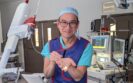
Kelvin Lau (pictured), consultant thoracic surgeon for St Bartholomew’s Hospital, said: ’The UK is leading the world in rolling out a national lung cancer screening programme. However, only some of the lung nodules identified during screening are cancerous and need treatment. Current biopsy techniques carry risk and are not always accurate, and many patients end up waiting for a repeat scan. The uncertainty of the wait causes anxiety and could allow a cancer to grow and spread.
’With this shape-sensing robotic technology, I have the precision and stability to lock onto a very small lung nodule and obtain an accurate biopsy quickly and safely. This could transform early diagnosis and treatment, reduce the need for repeat scans and treat lung cancer earlier.’
Also commenting on the ’transformative’ technology, Professor Pallav Shah, consultant respiratory physician based at Royal Brompton Hospital, added: ’We know that an earlier diagnosis of lung cancer leads to significantly improved outcomes for our patients. When we see patients with cancerous lung nodules of more than 30mm, their five year survival rate is around 68%, but if we are able to detect these nodules at a smaller size, when they are less than 10mm in size, we are looking at a 92% survival rate.’
There are already over 400 Ion systems installed in US hospitals, and Intuitive continues to explore its applications in other countries including in the UK, across Europe and beyond.
3rd August 2023
The use of magnetic tentacles offer a novel therapeutic and targeted approach for minimally invasive lung cancer detection and treatment, according to researchers from the University of Leeds.
The researchers developed a tiny, magnetically operated robot that is capable of travelling deep into the lungs and able to detect and treat the first signs of cancer. Their approach makes use of a 2.4 mm diameter, ultra-soft, patient-specific magnetic catheter – or tentacle – which can be delivered from the end of a standard bronchoscope to reach the periphery of the lungs. In addition, the tentacles possesses a laser fibre designed to enable targeted photo-thermal therapy to cancer cells.
In their study, which was published in Nature Engineering Communications, the team initially developed a three dimensional model of the bronchial tree, down to the sub-segmental bronchi, using data generated from a CT scan of the full lung. Once the tentacle was in position, laser light was delivered through the embedded fibre to induce thermal ablation of the tumour.
Following this initial and successful virtual experiment, the researchers next used the magnetic tentacle robot on the lungs of a cadaver. They were able to successfully navigate in three branches of the left bronchi, compared to only two using a standard catheter, which corresponded to a mean improvement in navigation depth of 37%.
Commenting on the results, Professor Pietro Valdastri, the project‘s research supervisor, said: ‘This is a really exciting development. This new approach has the advantage of being specific to the anatomy, softer than the anatomy and fully shape-controllable via magnetics. These three main features have the potential to revolutionise navigation inside the body.‘
Lung cancer has the highest worldwide cancer mortality rate. In early-stage non-small cell lung cancer, which accounts for around 84% of lung cancer cases, surgical intervention is the standard of care. In addition to being able to navigate within the lungs during a biopsy, the magnetic tentacle robot could pave the way for far less invasive treatment, allowing clinicians to target only cancer cells while allowing healthy tissue and organs to continue normal function.
Dr Giovanni Pittiglio, who carried out the research as part of his PhD, added: ‘Our goal was, and is, to bring curative aid with minimal pain for the patient. Remote magnetic actuation enabled us to do this using ultra-soft tentacles which can reach deeper, while shaping to the anatomy and reducing trauma.‘
26th June 2023
All smokers and ex-smokers aged 55-74 will have their risk of cancer assessed in the England’s first-ever national lung cancer screening programme.
The programme will be based on the Targeted Lung Health Check (TLHC) programme, which has been piloted in parts of England.
Under the plans, which will cost £270m annually once fully implemented, GP records will be used to identify patients for screening.
The first phase of the lung cancer screening scheme will reach 40% of the eligible population by March 2025, with the aim of 100% coverage by March 2030, the Government’s announcement said.
Patients will have their risk of cancer assessed based on their smoking history and other factors and those considered high risk will be invited for specialist scans every two years.
It is estimated the rollout will mean 325,000 people will be eligible for a first scan each year with 992,000 scans expected per year in total.
The UK National Screening Committee recommended in November that all four nations in the UK should implement a national lung cancer screening programme.
It said the TLHC programme would be a ‘practical starting point’ for implementation in England while a UK-wide programme needed ‘more modelling’.
During the pilots, approximately 70% of the screening took place in mobile units to ‘ensure easy access’ and ‘focused on more deprived areas where people are four times more likely to smoke’.
Almost 900,000 people were invited for checks, 375,000 risk assessments made and 200,000 scans were carried out.
Of these, more than 2,000 people were detected as having lung cancer, with 76% identified at an earlier stage compared to 29% identified outside of the pilot programme in 2019.
Urging patients receiving an invitation for lung cancer screening to go to their GP and take it up, NHS chief executive Amanda Pritchard said: ‘The NHS lung trucks programme is already delivering life-changing results, with people living in the most deprived areas now more likely to be diagnosed at an earlier stage, giving them a better chance of successful treatment.’
Health secretary Steve Barclay said: ‘Through our [lung cancer] screening programme we are now seeing more diagnoses at stage 1 and stage 2 in the most deprived communities, which is both a positive step and a practical example of how we are reducing health inequalities.
‘Rolling this out further will prolong lives by catching cancer earlier and reducing the levels of treatment required not just benefiting the patient but others waiting for treatment.
‘I am determined to combat cancer on all fronts through better prevention, detection, treatment and research.‘
Cancer Research UK’s chief executive, Michelle Mitchell, said: ‘This is really positive news for a cancer type that takes more lives than any other. Targeted lung screening across England could diagnose people most at risk at an earlier stage, when treatment is more likely to be successful.
‘For the screening programme to succeed, the UK Government must ensure that sufficient diagnostic equipment and staff are in place – a comprehensive and fully-funded NHS workforce plan for England will be vital to this.
‘Given smoking is the leading cause of lung cancer, it’s good to see that smoking cessation will be part of the programme. This needs to be embedded across all sites and stop smoking services must be properly funded to ensure people can quit smoking for good.
‘Other UK nations now need to follow suit to ensure everyone eligible can benefit from these potentially lifesaving lung checks.‘
A version of this story was originally published by our sister publication Pulse.
IMPORTANT LINKS
MORE FROM COGORA
© Cogora 2025
Cogora Limited. 1 Giltspur Street, London EC1A 9DD
Registered in the United Kingdom. Reg. No. 2147432