This website is intended for healthcare professionals only.
Take a look at a selection of our recent media coverage:
22nd December 2014
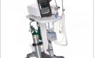
Royal Philips has announced the European launch of the V680 ventilator for hospital respiratory care, offering both invasive and non-invasive ventilation.
The solution is designed for smoother transition from ventilation to natural breathing, and helps enhance patient care and minimise ventilator associated infection rates. The V680 ventilator joins Philips’ existing portfolio of patient-centred hospital respiratory care solutions designed in consultation with clinicians, to deliver higher quality and comfortable care at lower cost.
“To ensure optimum respiratory care and a reduction in asynchrony, ventilators must be safe, effective, easy to apply and adapt to a patients’ changing condition”, commented Professor F. Javier Belda, Head of the Department of Anaesthesia and Critical Care, University of Valencia, Spain. “The V680 offers integrated invasive and noninvasive ventilation modes as well as flexible mask options. This helps my team facilitate quick therapy transitions and keeps our patients as comfortable as possible – while its clinically proven safety record is reassuring.”
“In respiratory healthcare we strive to advance our technologies to improve patient comfort and care beyond traditional mechanical ventilation”, says Arne Cohrs, Sales and Marketing Director, Therapeutic Care, Patient Care and Monitoring Solutions, Philips Healthcare EMEA. “The V680 ventilator has been developed in partnership with physicians, leading to customised technology that can be simply and quickly adjusted to respond to patient needs. Our goal is to speed the time to non-invasive ventilation, ultimately reducing hospital stays and associated cost burden.”
Non-invasive ventilation (NIV) has become a standard of care for the management of acute respiratory failure, but there is a risk of leaks around the mask that may interfere with ventilator performance. As a result, patient-ventilator asynchrony, defined as a mismatch between patient’s inspiratory time and the ventilator insufflation time, occurs in nearly 25% of intubated patients.1 These high rates of asynchrony are associated with a higher incidence of weaning failure and tracheostomy, prolonged mechanical ventilation, fatigue, increased sedation needs and longer hospital stays.1
A recent study to compare patient-ventilator synchrony during NIV between ICU, transport and dedicated NIV ventilators, found the use of a NIV ventilator on critically ill patients led to a significant decrease in the incidence of patient-ventilator asynchrony.2 Moreover, dedicated NIV ventilators exhibited more standardised behaviour during the investigation, with an ability to avoid auto-triggering or delayed cycling while keeping a short triggering delay despite the presence of leaks.2
The Philips V680 Ventilator will be available in several European markets by early 2015.
References:
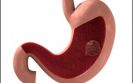
Eli Lilly and Company has announced that the European Commission has granted marketing authorisation for ramucirumab (Cyramza®).
Ramucirumab is the first licensed therapy specifically indicated for adult patients with advanced gastric (stomach) or gastro-oesophageal junction adenocarcinoma (GOJ), following prior chemotherapy (fluoropyrimidine and platinum). Ramucirumab is approved in combination with paclitaxel chemotherapy and as a single agent in patients for whom treatment with paclitaxel is not appropriate. Ramucirumab’s approval is based on two international Phase III studies, showing it extended overall survival time and delayed disease progression versus each study comparator.1–3
“Unfortunately, most patients with gastric cancer face a poor prognosis. Around 80% are first diagnosed once their cancer has spread and become difficult to treat. Despite research efforts, there have been few advances in the last 30 years and inoperable gastric cancer remains a devastating disease. Ramucirumab provides a welcome new treatment option for these patients,” says Professor David Ferry, Global Senior Medical Director, Lilly Oncology and previous Professor of Medical Oncology, New Cross Hospital, Wolverhampton, UK
Ramucirumab’s Phase III study programme involved 1,020 patients from 289 cancer centres. Results from the RAINBOW study showed that ramucirumab, combined with paclitaxel, significantly extended overall survival time to 9.6 months compared with 7.4 months for placebo plus paclitaxel (hazard ratio [HR] 0.807 [95% CI 0.678–0.962]; p=0.017).2
As a single agent in the REGARD study, results showed ramucirumab extended overall survival time to 5.2 months compared with 3.8 months for best supportive care (HR 0.776, 95% CI 0.603–0.998; p=0.047).3 In both studies, ramucirumab was shown to delay disease progression and improve objective response rate ie tumour shrinkage.2,3
In the combination study, the most common adverse events of grade 3 or higher for ramucirmab plus paclitaxel versus placebo plus paclitaxel were neutropenia (41% vs 19%), leucopenia (17% vs 7%), hypertension (14% vs 2%), fatigue (12% vs 5%), anaemia (9% vs 10%) and abdominal pain (6% vs 3%). In the single agent study, adverse events were mostly similar between the ramucirumab and placebo groups, although rates of hypertension were higher (16% vs 8%).
‘Lilly is proud to have developed the first licensed therapy specifically indicated for second-line advanced stomach cancer and gastro-oesophageal junction adenocarcinoma. Today marks an important milestone for patients with this difficult-to-treat disease. Ramucirumab is an innovative therapy that specifically binds to and blocks vascular endothelial growth factor (VEGF) Receptor 2 – a key mediator of VEGF-induced angiogenesis, which feeds cancer growth. Ramucirumab’s development is testament to our commitment to support people living with cancer and those who care for them’ said Richard Gaynor, MD, Senior Vice President, Product Development and Medical Affairs for Lilly Oncology.
Ramucirumab was granted Orphan Drug Designation by the European Commission for treatment of gastric cancer in the EU. Orphan drug status is designated to medicines for the treatment of rare diseases, where available options are limited or where a medicine offers significant benefit over existing treatments.
References
Ipswich Hospital NHS Trust, (IHT) which provides healthcare services to 356,000 people across Ipswich and East Suffolk, has revealed that it is using location analytics solutions from leading B2B mapping and analytics company, Esri UK.
IHT is making use of the solutions as part of a feasibility programme to assess how location analysis and actuarial analysis working together can provide insight into demand management to shape future hospital services for the better.
IHT recognises the huge potential of the approach to achieve faster time to insight. Visualising data in a map format helps IHT understand where the hotspots are for certain diseases, admission methods and average time to treatment. Understanding where demand comes from into emergency admissions, and the type and level of that demand, has allowed IHT to pinpoint areas of intervention to mitigate it.
The hospital believes this will enable it to improve its resourcing processes. It will be able to bring in more clinical support staff to meet increased demand on certain days of the week, for example, and therefore alleviate resource pressure. This should in turn help minimise costs while maintaining performance and clinical standards.
Paul Scott, director of finance and performance at IHT, says: “While this remains an R&D project it has highlighted a different way of looking at our organisation, and the demand pressures we face. In addition, it has demonstrated the huge potential of looking at demand from a population rather than individual attendance basis.
“It also allows us to try out ‘what if’ scenarios. In short, it has given us a whole new platform for discussion and changed the conversation at board level to focus on moving to a more sophisticated way of visualising and reporting data. The IHT board has even requested that geographic information and location is included within its business intelligence strategy.”
The solution provides IHT with a combination of insight from Esri’s geographical information systems (GIS) and location analytics technology and high-quality clinical and financial data analysis, feeding into long-term financial modelling, provided by the actuarial service provider.
Esri UK’s location analytics solution was vitally important in delivering data visualisation to provide the required insight. Utilising the ArcGIS platform on IHT’s infrastructure behind a secure firewall, ArcGIS for Server and ArcGIS for Desktop were deployed to provide services to a management insight dashboard.
“The hospital has gained additional insight into demand for its A&E services by using our solution to map out A&E admissions by a range of variables,” says Dr Ed Wallington, health and social care lead, Esri UK. “By visualising the information as a hotspot analysis, it has been able to highlight the source of demand. And it has achieved a much greater understanding of the status of demand by using our Key Performance Indicator (KPI) reporting tools to break down A&E attendees by number of cases, average waiting time or mode of travel, and then split the figures down by timeframes.”
Moving forward, the trust believes the outputs from patient demand analysis, mapping insights and possible interventions could provide a basis for service reconfiguration and therefore wastage reductions. The use of GIS information, in particular in conjunction with actuarial, could put IHT ahead of other National Health Service Trusts in terms of innovative use of IT in healthcare provision.
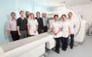
St George’s Healthcare NHS Trust has become the first in the UK to install the new Symbia Intevo™ xSPECT technology from Siemens Healthcare.
The Trust has installed two of the systems, a Symbia Intevo Excel and Symbia Intevo T16 as part of a major upgrade within its nuclear imaging diagnostic facility.
The Intevo technology is joined by a Symbia® S multi-purpose SPECT system with the three new installations helping to meet present and future workload requirements within the Trust.
The Symbia systems have replaced three gamma cameras within the department and are being used for a wide range of procedures, including tumour, infection and skeletal imaging. The xSPECT technology integrates high sensitivity single-photon emission computed tomography (SPECT) and high specificity CT, combining them to provide anatomical and functional information. The excellent image quality and additional available information has improved diagnostic confidence to clinicians in the department.
“We are delighted to be the first site to install the latest Intevo technology from Siemens Healthcare,” states Andy Irwin, Consultant Clinical Scientist at St George’s Healthcare NHS Trust. “We have completely revitalised the department, and Siemens were on hand to help with the design of a joint control room, enabling us to continue to increase efficiency and departmental productivity. The team are very happy with the applications training received and have found the interface across all three Symbia systems very intuitive.”
The new Symbia Intevo xSPECT systems reconstruct both the SPECT and CT portions of an image into a high frame of reference for precise, accurate alignment of information allowing for differentiation between tissue boundaries in bone imaging. The systems use more CT data than ever before and are still able to limit patient dose by offering Combined Applications to Reduce Exposure (CARE). The systems also use AUTOFORM collimator, capturing up to 26% more counts, helping boost image acquisition time and patient throughput.
Malcolm Pickering, Regional Sales Manager at Siemens Healthcare states, “The Symbia Intevo is the world’s first xSPECT system and demonstrates Siemens’ commitment to continuous innovation. The SPECT and two xSPECT systems are a major upgrade to St George’s Nuclear Medicine Department and are already providing clinicians with additional confidence when it comes to supplying an accurate and rapid diagnosis for patients.”
17th December 2014
A satellite symposium entitled Managed equipment services: together, better, stronger? was presented at the HOPE Congress 2014 on 27 May in Amsterdam.
The presentation was given by Mr Lodewijk Wuite, General Manager MES, Toshiba Medical Systems Europe BV. The purpose of the Symposium was to illustrate the benefits to hospitals of a managed equipment service.
In healthcare, many countries around the world are confronted with an ageing population, smaller workforces to support them, assertive patients, and increases in chronic and lifestyle-related diseases, such as COPD, obesity, and cancers, to name but a few, creating an increased demand for healthcare. By contrast, budgets are being decreased continuously, best practices are not very well shared, production is still favoured over quality, and e-health harmonisation is only making slow progress. In short, healthcare systems will become unsustainable and unaffordable in the near future if no major breakthrough occurs.
In May 2014, COCIR, the European Coordination Committee of the Radiological, Electromedical and Healthcare IT Industry, published a study that presented alarming data related to ageing equipment (Table 1): with the exception of a few countries (typically those countries that received structural funds for investments in healthcare from the EU in the last years), the average age of high technology equipment is increasing. In all computed tomography, magnetic resonance imaging and angiographic cardiovascular systems, the percentage of installed base of one to five years old is decreasing. This means that, even though we have become more demanding and we want to have better images, faster procedures and newer technologies, in reality, there is an issue with the age of the equipment: it cannot keep up with the demands of the market.
___________________________________________________
BOX 1
Toshiba Corporation, a Fortune Global 500 company, channels world-class capabilities in advanced electronic and electrical products and systems into five strategic business domains: energy and infrastructure; community solutions; healthcare systems and services; electronic devices and components; and lifestyle products and services. Guided by the core principles of ‘Committed to People, Committed to the Future’, Toshiba promotes global operations towards securing ‘Growth Through Creativity and Innovation’, and is contributing to the achievement of a world in which people everywhere live in safe, secure and comfortable society.
Founded in Tokyo in 1875, today’s Toshiba is at the heart of a global network of over 590 consolidated companies employing over 200,000 people worldwide, with annual sales surpassing 6.5 trillion yen (US$63 billion).
___________________________________________________
What are managed equipment services?
The concept of managed equipment services (MES) is the outsourcing of all aspects of medical technology to a third party company that specialises in providing this type of service. This company will provide the expertise to purchase, install, replace, manage and maintain a portfolio of medical equipment, and train users on a long-term basis. The MES provider will usually own the equipment and provide it as part of a managed service, which encompasses all the necessary elements to support effective use of the equipment. Because the MES provider has a single focus on medical equipment and a larger presence in the market than individual healthcare providers, it is able to provide a higher standard of service at a cost-effective price.
The outsourcing of non-core activities is common in large organisations that seek to transfer risk to third-party specialist organisations that are better able to manage it. This can enable the healthcare provider management to concentrate on its core role of providing a high standard of clinical services to patients.
MES provide a strategic approach to managing technology, which ensures that medical equipment is maintained to a high standard and is replaced through an agreed investment plan. The replacement plan is flexible over time and allows for modifications in cases of hospital strategic or environmental change. This ensures that patients and clinicians always have access to the required standard of equipment to perform their work, thereby reducing clinical risk and increasing productivity. With MES in place, the availability of medical equipment is rarely a concern.
The need for a MES
The role of the industry is changing in the healthcare arena. Companies should support hospitals in achieving their strategic and operational objectives, thereby creating a state of togetherness (proactive collaboration and co-creation), that is better (leading instead of suffering from changing circumstances) and stronger (transferred/shared financial operational and technological risk, and shared responsibility and success). One of the tools that can help to achieve this is a MES.
What is a MES?
At its core, it is the supply of functionality rather than the sale and service of equipment, under a long-term outsourcing contract, with the aim of:
A MES is hence not just a financial programme whereby a piece of equipment is leased. It is about:
A conceptual overview of a MES programme is indicated in the flowchart in Figure 1, while typical added values are shown in Figure 2.
Contracting
The plan of action once a contract is signed include:
During the contract, improvement actions will be defined, developed and executed. Technology will be adjusted where needed.
Financial calm
From a cash flow and balance sheet perspective, the hospital moves from a situation of varied capital spend, requiring repeat business cases, to a situation of known fixed monthly charges, with off-balance sheet financing (Figure 3).
For financial management of change, hospitals can follow their own MES project and simulate changes via Toshiba’s proprietary In-the-Cloud Amp2hi® software (Figure 4).
This software supports financial management of a MES contract. It allows calculations of monthly fees. It is also capable of managing change of contract contents in all its aspects and the effects this has on the fee during the contract. Customers are able to access their project through the internet and simulate changes in technology and/or services compared with the contractual content and the effects on the monthly fees.
Access to innovation
Innovation cycles are much shorter than economic life cycles. What does that mean? Often, when a hospital buys a new piece of equipment and the radiologists, or other specialists, have an input on what they want, they focus on latest state of the art. The rationale being that if they have to work for a long, accounting-based, period of time with a system, they know that after a couple of years the technology might become obsolete. In order to cope with this, they want to see this point as far as possible in the future and consequently procure as high specified system as possible.
That means that a hospital would need to invest more money than required based on the actual functional requirements. By running a managed service, and by sometimes bringing in lower class, new systems but replacing them much more rapidly, or bringing high class refurbished systems, hospitals can remain continuously within the clinical and technological requirements of the departments. Of course, sometimes state of the art technology is required, for example if there are R&D programmes initiated and/or ongoing. But in daily practice, state of the art is not always required. It is most often sufficient to work with state of need technology.
State of need is about bringing the exact technology that you require.
This is well illustrated in the following models of functionality versus investment: the former (Figure 5), the traditional model, and the latter (Figure 6) showing how MES counters the pitfalls, to keep within the targeted functionality range.
Critically, MES assures like-for-like functionality when equipment is replaced in the course of the contract, whereby clinical freedom of choice should be provided.
There are many and varied optional services, depending on the customer’s requirements, some of which include:
Summary
A managed service is a contractual partnership between a healthcare provider and a technology provider typically for a period of about 10–15 years. In this period, it will replace all of the equipment. It facilitates the supply and maintenance of equipment; it can include financing as well. It takes care to ensure that all the help and first-line service is streamlined for the involved hospital’s imaging departments.
It provides continuous education and training. Besides that, MES provides a variety of additional improvement programmes, that are developed and executed in close collaboration with hospitals.
Another key for a managed service is that there is no technological overkill or underkill. That means that the functionality that is needed at any point in time, taking into consideration changes in environment and requirements, is supplied. And importantly, clinical freedom of choice is guaranteed.
16th December 2014
Aseptix Health Sciences NV, the specialist in next generation antimicrobials headquartered in the Netherlands, announced a global licence agreement with Ecolab.
Under the agreement, Ecolab will acquire a global licence for Professional Markets on Aseptix technologies and acquire the Professional Products Division of Aseptix. Aseptix currently sells its Aseptix-branded professional products in Europe and India, mainly to healthcare, food processing and pharmaceutical clients.
Aseptix is the inventor of activated hydrogen peroxide technologies, a fast-acting and user-safe technology to effectively eradicate numerous pathogens with outstanding material compatibility and without burdening residues. The active ingredient degrades to water and oxygen after use.
Under this transaction, Aseptix will continue to own the intellectual property of more than 60 patents around its technologies and related know-how, while Ecolab will receive exclusive rights for use of the majority of Aseptix’s technologies in professional markets around the world. This enables Aseptix to stay independent and capitalise on its proven technologies for consumer applications across many markets globally.
The agreement with Ecolab fits the Aseptix strategy of globalising the technology and creating the new global standard in infection prevention. With the growth in hospital acquired infections (HAI) and increasing focus on clean environments, the relevance of these types of solutions is rapidly increasing.
It is Aseptix’ ambition to offer state of the art products in the fight against dangerous microbes. The Aseptix technology is unique, since it offers broad efficacy spectrum and short contact times, is safer for people and environment and is very easy to use.
Ilja Bobbert, founder and CEO of Aseptix: “This is a major milestone for Aseptix. Aseptix has laid the initial groundwork and has proven the products can lower infection rates in many industries. Now, Ecolab can truly globalise the technology across their business segments, and geographies worldwide.”
For many years, Aseptix is a trusted partner with a proven track record in building long lasting and profitable partnerships with market leaders in various industries across the globe. The company’s objective is to exploit Aseptix’s safe and environmentally friendly technologies in numerous antimicrobial applications globally. With this transaction aimed at the professional markets, Aseptix can improve its company efforts on licencing the technologies to household care and personal care applications.
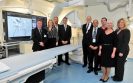
Basingstoke and North Hampshire Hospital has officially opened a newly refurbished Interventional Radiology Theatre that now incorporates a state-of-the-art angiography unit, the Artis zee™ ceiling-mounted system from Siemens Healthcare.
The installation of this system will provide the hospital, part of Hampshire Hospitals NHS Foundation Trust, with the ability to expand and enhance oncology services to patients.
The Artis zee ceiling-mounted system incorporates a range of advanced applications such as syngo® DynaCT, soft-tissue 3D imaging, and iGuide live integrated needle guidance. This functionality enables users at the hospital to plot a direct path to organs for biopsies and drainages within the Interventional suite with increased speed and accuracy. The use of syngo Embolisation Guidance supports oncology procedures with the ability to directly target the blood supply specific to tumours and deliver chemotherapy beads for treatment.
The new system was unveiled at an official opening for the new Interventional Radiology Theatre, attended by hospital Board members and Directors alongside senior representatives from Siemens Healthcare. Dr Graham Plant, a retired Consultant Radiologist at the hospital, and Wendy James, a semi-retired Superintendent Radiographer, who have been pioneers of interventional radiology over the past 30 years, were responsible for the unveiling.
The system’s advanced software capabilities provide the ability to merge previous CT and MR scans with live images in real time. This opens a new world of treatment options with confidence through the use of 3D X-ray datasets. The system, which offers a comprehensive Sensis and cardiac package, has provided the hospital with the ability to perform advanced cardiac device implantation.
Dr Gaurang Ubhayakar, Interventional Radiologist at Basingstoke and North Hampshire Hospital, states, “CT scanners have significant pressures placed on them in terms of providing timely diagnoses, and this new system will assist with needle-guided microwave and radiofrequency ablation of tumours that are time consuming on the CT scanner, amongst other procedures. Our new system transfers CT guided interventional procedures, thereby increasing CT diagnostic capacity.”
“Developed specifically for interventional radiology, the Artis zee‘s large flat detector affords easy patient access and full body coverage, with the system’s dual functionality combining Angiography and Cardiology together within one solution,” states Paul Vaughan, Regional Sales Manager at Siemens Healthcare. “We are pleased to be able to support Basingstoke and North Hampshire Hospital’s important work in this area.”
Siemens Healthcare is one of the world’s largest suppliers to the healthcare industry and a trendsetter in medical imaging, laboratory diagnostics, medical information technology and hearing aids. Siemens offers its customers products and solutions for the entire range of patient care from a single source – from prevention and early detection to diagnosis, and on to treatment and aftercare. By optimising clinical workflows for the most common diseases, Siemens also makes healthcare faster, better and more cost-effective. Siemens Healthcare employs some 52,000 employees worldwide and operates around the world www.siemens.co.uk/healthcare.
15th December 2014
Bare metal stent (BMS) usage continues to decline. However, there are still conditions when a BMS should be used. In such cases it is important to use the BMS with the lowest target lesion revascularisation (TLR) rate. The REBEL™ stent system, from Boston Scientific, is the newest advance in BMS technology – its advantages include radial strength and low recoil of stent, as TLR rates are generally higher for a BMS compared with a drug-eluting stent.
Ieva Briede
Andrejs Erglis PhD FESC FACC
Pauls Stradins Clinical University Hospital, Latvian Cardiology Center andPauls Stradins Clinical University Hospital, University of Latvia, Latvia
Key words: Stents; imaging; coronary disease
Date received: 20 July 2014
Date accepted: 7 August 2014
For decades, the main limitation of percutaneous treatment of coronary lesions has been restenosis, which affects 10% to 30% of patients. After plain balloon angioplasty, restenosis is mainly due to elastic recoil and negative remodelling of the vessel wall. Intravascular implantation of a metallic stent prevents both problems and results in less restenosis compared with balloon angioplasty alone. However, stent implantation induces a foreign body reaction, leading to excessive neointima formation, which in turn causes in-stent restenosis.1 Drug-eluting stents (DES) reduce neointima formation, but at the expense of a prolonged risk of very late thrombosis due to delayed endothelialisation, earlier and more frequent neoatherosclerosis or both. Data on second-generation DES in combination with prolonged dual antiplatelet therapy are more reassuring on late thrombosis but are currently limited to two to four years follow-up for very late thrombosis.2,3 There has been tremendous progress in stent technology from bare metal stents (BMS) to DES and to biodegradable polymer DES. DES revolutionised the practice of interventional cardiology and coronary revascularisation with significant reductions in the risk of restenosis and consequently the need for repeat revascularisation compared with BMS.4,5 However, despite the introduction of DES more than ten years ago, BMS are still being used, although with varying frequency.6
The use of DES and BMS
The European Society of Cardiology (ESC) guidelines on myocardial revascularisation in 2010 recommended DES usage in almost all patients, but there were some relative clinical contraindications to the use of DES and there was some place for BMS (Table 1).
Recently, Bangalore et al evaluated the trend in DES use across the US from 2001 to 2011.8 Among the 8.1 million coronary procedures, drug-eluting stent use reached a peak in 2005 at 89% in all patients including groups with a low risk of restenosis, high risk of stent thrombosis or bleeding. A steep drop to 66% was noted in 2007 followed by a progressive rise to 73% in 2011, when BMS were used in 26% of all stent implantation. Compared with 2001, the adjusted odds ratio of BMS use dropped 95% by 2011. In 2011, 75% patients with diabetes, 66% patients with a history of bleeding peptic ulcer and 61% patients with a history of atrial fibrillation received DES. BMS was still often used in elderly patients, patients with peptic/gastric ulcers, patients with planned surgery and in those who required long-term anticoagulant therapy.
Although DES usage is increasing in the US, there are some conditions when BMS should be used. In such cases it is important to use the BMS with the lowest target lesion revascularisation (TLR) rate.
BMS studies in the DES era
Many studies have been performed to evaluate the ‘best’ BMS and a number of alternative approaches to DES have been tested. The Genous bio-engineered BMS carries a layer of murine, monoclonal, antihuman CD34 antibody, aimed at capturing circulating endothelial CD34+ progenitor cells, thus possibly increasing the rate of healing. The single-centre pilot TRI-stent adjudication study (TRIAS) did not confirm initial promising results in patients at high risk of coronary restenosis.9 Another single-centre observational study comparing silicon carbide-coated metal stents (PRO-Kinetic) and uncoated BMS (Vision) in 2731 patients was performed in Finland. The PRO-Kinetic stent remained an independent predictor for revascularisation (p=0.002). This study showed that silicon carbide-coated BMS was associated with greater target lesion revascularisation rates at one year compared with the Vision stent.6
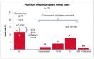
The OMEGA study was the latest prospective, multicentre, single-arm study, which enrolled 328 patients at 37 investigative sites in the US and the EU. Patients received the OMEGA(™) stent (the new REBELTM is the same platinum chromium platform of OMEGA(™), but with innovative customised stent architecture) for the treatment of de novo native coronary artery lesions (≤28mm long; diameter ≥2.25mm to ≤4.50mm). The primary endpoint was nine-month target lesion failure (TLF: cardiac death, target vessel-related myocardial infarction, TLR) compared with a prespecified performance goal based on prior-generation BMS. All major cardiac events were independently adjudicated. Dual antiplatelet therapy was required for a minimum of one month post procedure. Nine-month outcomes of the OMEGA study showed a low rate of TLF, revascularisation and stent thrombosis. The primary endpoint was met – the nine-month TLF rate was 11.5% and the upper one-sided 95% confidence bound of 14.84% was less than the prespecified performance goal of 21.2% (p<0.0001). Event rates were low, including a stent thrombosis rate of 0.6% at nine months. Through nine months, the myocardial infarction rate was 3.7% (12/326). All of the myocardial infarctions were non-Q wave myocardial infarctions, with most occurring within one day of the procedure (10/12). This supports safety and efficacy of the novel, platinum chromium OMEGA/REBEL BMS for the treatment of coronary artery disease (Figure 1).10
The REBEL(™) stent
The REBEL™ stent system is the newest advance in BMS technology. Radial strength and low recoil of stent are particularly important, as TLR rates are generally higher for BMS compared with DES. The platinum chromium alloy combined with the REBEL™ customised stent architecture allows for high radial and axial strength and the lowest stent recoil. REBEL™ is based on the OMEGA™ stent, but utilises a slightly modified stent platform with additional connectors added to the proximal end of the stent to reduce the potential for stent deformation. In bench testing, the REBEL™ stent outperforms its competitors in deliverability tests such as trackability and pushability, which are two important requirements for physicians when delivering a BMS. Platinum chromium alloy gives the best-in-class visibility during coronary stenting (if comparing with other BMS platforms). Platinum has over twice the density of iron or cobalt, resulting in enhanced visibility under fluoroscopy.11
REBEL has another useful feature: the post-dilatation overexpansion limit to 5.75mm for the large vessel stent model, resulting in optimal post-dilatation capability for big vessels.
Optimal implantation technique
Although DES have superior outcomes to BMS in terms of both restenosis and stent thrombosis, we have to agree that there is still a place for BMS in modern catheterisation laboratory. Moreover, with the optimal stent implantation technique we can achieve less TLR and stent thrombosis in both BMS and DES systems.
Colombo et al has shown that when using intravascular ultrasound, the majority of stents are not adequately deployed, despite a successful angiographic result. It was therefore hypothesised that the major cause of stent thrombosis was incomplete expansion rather than intrinsic thrombogenic properties of the device.12
In 1998, De Jaegere et al validated the safety and feasibility of intravascular ultrasound-guided stenting and its impact on the six-month restenosis rate in the Multicenter Ultrasound Stenting in Coronaries (MUSIC) study.13 They confirmed that, in selected patients, stents could safely be implanted without the use of systemic anticoagulation, when optimal stent expansion was achieved (Table 2). Of all variables predictive of stent thrombosis, the final angiographic result reflecting the degree of stent expansion has consistently been shown to be one of the most important predictors. These data corroborate previous studies in which it was found that the lumen diameter immediately after the procedure was one of the most powerful independent predictors of coronary patency at six months.
However, the optimal implantation technique and adjunctive pharmacological treatment still need to be established. The risks from aggressive stent expansion are coronary rupture or extensive vessel wall injury, which in turn may provoke a severe vessel wall response and thus restenosis. Appropriately sized balloons based on intravascular ultrasound measurements can avoid this. This may, in part, explain the low restenosis rate observed in the MUSIC study.
Conclusions
A significant proportion of patients still receive BMS. From many BMS types, the operator should use the BMS with the best performance and the lowest rate of TLR. The optimal stent implantation technique including postdilation with non-compliant balloon is compulsory. Intravascular ultrasound can help to choose right diameter stent and balloon size. As well as right lesion pretreatment, predilatation with balloons, cutting/scoring balloons, rotablator, etc, is mandatory for optimal stent implantation.
Key points
References
12th December 2014
St. Jude Medical, Inc announced CE Mark approval of the Quadra Allure MP™ cardiac resynchronisation therapy pacemaker (CRT-P).
The Quadra Allure MP is the world’s first and only quadripolar CRT-P with the MultiPoint Pacing option, a technology that has been shown to enhance patients’ response to CRT, potentially improving quality of life for patients with heart failure.
The MultiPoint Pacing technology enables physicians to pace multiple locations on the left side of the heart, giving the clinician more choices to best optimise CRT pacing based on patient need and reducing the rate of CRT non-responders, as well as the likelihood of costly and invasive lead revision through a second intervention procedure.
A recent study demonstrates that MultiPoint Pacing technology may be particularly beneficial in patients not responding to traditional bi-ventricular pacing therapy, which accounts for approximately one third of the total population of patients receiving cardiac resynchronisation therapy (CRT). The technology, developed by St. Jude Medical, has demonstrated a 19% improvement in responder rates for patients with MultiPoint Pacing at 12 months compared to traditional methods of CRT. Additionally, research found improvement in left-ventricular (LV) function in patients who were already classified as a responder to CRT therapy.
The Quadra Allure MP CRT-P is designed to work with the Quartet™ LV lead, which has 4 electrodes to offer maximum flexibility for different pacing configurations to help manage heart failure patients. The MultiPoint Pacing capability allows physicians to program simultaneous or sequential delivery of two LV pulses at 2 different anatomical locations per pacing cycle, rather than the standard single pacing pulse, which can result in more effective resynchronisation, potentially leading to better clinical outcomes compared to single site pacing.
St. Jude Medical developed the quadripolar technology, launching the first ever quadripolar lead in 2011 and entered the pacemaker market in April 2013 with the Allure Quadra™ CRT-P System. Quadripolar technology offers physicians more options to manage heart failure and facilitates additional pacing configurations within the heart that offer physicians options not available in traditional bipolar systems. Earlier this year, the MORE-CRT prospective, randomised clinical trial of more than 1000 patients demonstrated a 40% relative risk reduction of lead-related complications for patients implanted with the Quartet LV lead. To date, more than 100 clinical publications have provided broad clinical evidence supporting the advantages of quadripolar technology from St. Jude Medical.
The announcement follows data that was presented during the XVI International Symposium on Progress in Clinical Pacing in Rome. Building upon the 19% improved responder rate, data shows that MultiPoint Pacing improved LV function for both responders and non-responders compared to traditional CRT. Additionally, a study suggests that multipoint LV pacing may reduce the burden of premature ventricular contractions (PVC), potentially improving the effectiveness of CRT.
“From our initial experience, we think MultiPoint Pacing improves cardiac function, resulting in better resynchronisation and could be a further advantage of CRT,” said study investigator, Dr. Francesco Zanon, MD, FESC, FHRS, director of the Electrophysiology Unit Department of Cardiology from Santa Maria della Misericordia Hospital in Rovigo, Italy. “We observed significant improvement over traditional CRT pacing, and therefore believe this technology could have wide application because it is accessible for all CRT implanters.”
“The St. Jude Medical quadripolar pacing system has been successfully designed for optimised outcomes, now adding MultiPoint Pacing as an additional set of tools designed to decrease the rate of non-responders and improve clinical outcomes even in difficult to treat patients with ischemic heart disease,” said Eric S. Fain, MD, group president of St. Jude Medical. “Building upon the industry’s first quadripolar platform, the MultiPoint Pacing technology is an excellent example of our continued commitment to investing in innovative solutions that reduce health care costs and improve outcomes for patients.”
MultiPoint Pacing is an investigational device and is not commercially available in the US.
Quadripolar Pacing Technology from St. Jude Medical
St. Jude Medical introduced the industry’s quadripolar pacing system featuring four pacing electrodes. The quadripolar pacing system offers physicians the ability to effectively and efficiently manage the ever-changing needs of patients with heart failure. The system integrates multiple pacing configurations and Tailored Therapy™ features that enable physicians to optimise the system at implant and follow-up, as well as better manage common pacing complications without having to surgically reposition the lead.
The Quartet lead design allows the physician to implant the lead in the most stable position without making trade-offs in electrical performance. This includes pacing closer to the base of the left ventricle, which studies associate with better patient outcomes and which may be more difficult with traditional bipolar leads. The quadripolar pacing electrodes also provide physicians more options to optimise CRT performance, such as pacing around scar tissue in the heart and avoiding the most common pacing complications.
10th December 2014
Digital autopsies are set to become an increasingly common practice across parts of the UK with the opening of a number of new facilities providing the service.
The digital autopsy facilities will provide comfort to families, in many cases allowing a cause of death to be identified swiftly and with either no or minimal invasion of the body, which in turn can help make the burial or cremation process take place with fewer delays and ease the emotional burden.
iGene®, the lifescience company investing £50m in creating a network of digital autopsy centres across England and Wales, is expanding its service with the use of 6 SOMATOM® Definition AS CT systems from Siemens Healthcare. The company has recently installed the first of the imaging systems into its dedicated Sandwell facility in the West Midlands to support pathologists across the region in determining the cause of death. Following the Sandwell installation, the additional systems will be placed in various sites throughout the UK in the coming year.
It is predicted that digital autopsy could be used to establish the cause of death in up to 70% of cases and they are particularly valuable for identifying cerebral haemorrhage, lung pathology, fatal aneurysms and extent of trauma leading to fatality. Certain cases, which could be difficult to identify during a conventional post mortem, can also be more easily identified using the digital technique, often improving the accuracy of results. Where digital autopsy cannot provide the cause of death alone it can instead help to minimise the need for incisions to the body.
“The merging of innovative CT technology with specialist software will allow these centres to perform advanced examinations, providing a comprehensive picture of the cause of death without using the invasive techniques that we see in current post-mortem practice,” states Russell Lodge, CT Business Manager at Siemens Healthcare. “The systems will provide radiographers, radiologists and pathologists excellent image quality and sharp contrast to provide an accurate cause of death.”
“Post-mortem imaging is quite different to ante-mortem imaging and it requires the reverse of a lot of traditional scanning practices. Siemens understood what we were trying to achieve and were flexible, tailoring their service to our unique requirements,” states Claire Walker, Digital Autopsy Services Manager at iGene. “The new facilities offer not only comfort for families who are seeking to understand how their loved one died, in a swift and dignified manner, but a highly innovative option for authorities who can use them within investigations.”
The SOMATOM Definition AS systems were chosen due to the user friendly and intuitive interface and Siemens’ unique STRATONTM X-ray tube offering. The virtual post-mortem process acquires a whole body scan at 0.6mm, therefore a robust X-ray tube is required to avoid tube overheat. STRATON X-ray tube offers the combination of high speed and excellent image quality eliminating the need for heat storage capacity with the tube cooling down to its original state within 20 seconds. The systems work alongside iGene INFOPSY® visualisation software to provide either non- or minimally-invasive autopsies.
IMPORTANT LINKS
MORE FROM COGORA
© Cogora 2025
Cogora Limited. 1 Giltspur Street, London EC1A 9DD
Registered in the United Kingdom. Reg. No. 2147432