Dr Sumeet Hindocha has a passion for artificial intelligence, with his work focusing on radiomics and deep learning in lung cancer. He speaks to Hospital Healthcare Europe about his latest research and the uses and considerations of AI-enabled diagnostics in medicine.
Dr Sumeet Hindocha is a clinical oncology specialist registrar at The Royal Marsden NHS Foundation Trust and a researcher in artificial intelligence (AI). He is currently leading the trust’s OCTAPUS-AI study to investigate how this technology can help identify which patients with non-small cell lung cancer are at higher risk of recurrence.
Why are you interested in lung cancer and AI?
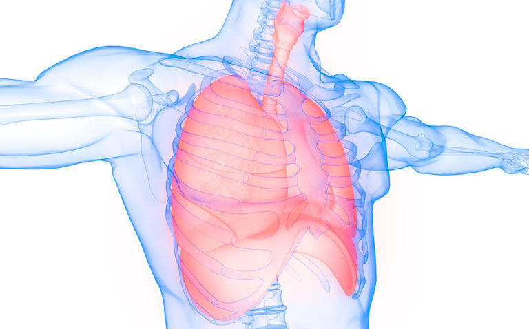
Lung cancer is the leading cause of cancer deaths worldwide. Non-small cell lung cancer (NSCLC) is behind almost 85% of cases and is often curable when detected early enough. Radiotherapy is a key treatment modality for it, but, unfortunately, recurrence can occur in over a third (36%) of patients treated with radiotherapy.
We know that the earlier we detect recurrence the better the outcomes generally are for patients. It means we can get them on to the next line of treatment or offer the best support as soon as possible. This could reduce the impact the disease has on their lives and help patients live longer.
The aim of our study is to see whether AI could help identify the risk of cancer returning in these patients using CT scans. The study addresses the National Institute of Healthcare and Clinical Excellence’s call for further research into using prognostic factors to develop risk-stratification models to inform optimal surveillance strategies after treatment for lung cancer.
Where does your enthusiasm for AI stem from?
Artificial intelligence has had a big impact in improving various aspects of our lives and work, from automating routine tasks to even things like the programmes recommended to us on Netflix or smart home devices like Siri or Alexa. What’s really exciting about its application in healthcare is its significant potential to improve patient outcomes and experience. We have a huge amount of data from imaging and electronic patient records that can be readily applied to AI. It gives us the ability to detect patterns of disease that would otherwise be difficult to uncover, to develop new drugs and even streamline how we deliver healthcare.
Who are you working with on the OCTAPUS-AI study?
Researchers from the Institute of Cancer Research, Imperial College London and the Early Diagnosis and Detection Centre, which aims to accelerate early diagnosis of cancer and is supported by funding from the Royal Marsden Cancer Charity and the National Institute for Health and Care Research.
What did the first phase of the study involve?
We compared different models of machine learning (ML) – a type of type of AI that enables computer software to learn complex data patterns and automatically predict outcomes – to determine which could most accurately identify NSCLC patients at risk of recurrence following curative radiotherapy.
Anonymised, routinely available clinical data from 657 NSCLC patients treated at five UK hospitals was used to compare different ML algorithms based on various prognostic factors such as age, gender and the tumour’s characteristics on scans to predict recurrence and survival at two years from their treatment. We then developed and tested models to categorise patients into low and high risk of recurrence, recurrence-free survival and overall survival.
A patient’s tumour size and stage, the type and intensity of radiotherapy, and their smoking status, BMI and age were the most important clinical factors in the final AI model’s algorithm for predicting patient outcomes.
The results suggested that this technology could be used to help personalise, and therefore improve, the surveillance of patients following treatment based on their risk. This could lead to recurrence being detected earlier in high-risk patients, ensuring that they receive urgent access to the next line of treatment that could potentially improve their outcomes.
Results from the second phase of the study were recently published. Can you tell us more about this work?
In this phase, as well as clinical data, we used imaging data describing the tumours’ characteristics – a technique known as radiomics – taken from radiotherapy treatment planning CT scans on over 900 NSCLC patients in the UK and Netherlands.
Radiomic data can also be linked with biological markers. We believe it could be a useful tool in both personalising medicine and improving post-treatment surveillance. This data was used to develop and test ML models to see how accurately they could predict recurrence.
The TNM staging system, which describes the amount and spread of cancer in a patient’s body, is the current gold standard in predicting prognosis. However, our model was found to better correctly identify which NSCLC patients were at a higher risk of recurrence within two years of completing radiotherapy than a model built on the TNM staging system.
How could your findings benefit patients?
We are at an early stage, and there’s a lot more work to do before we have a tool ready for use in the clinic. However, our results suggest that our AI model could be better at predicting tumour regrowth than traditional methods. This means that, using our technology, clinicians may eventually be able to identify which patients are at a higher risk of recurrence and offer them more targeted follow up. If recurrence did occur, this would be detected earlier so patients could be offered the next line of treatment as soon as possible. Meanwhile, low-risk patients could potentially be spared unnecessary follow-up scans and hospital visits.
This is also an exciting project because we don’t have to put patients through extra procedures for the model to work, as the data is routinely collected during the course of their normal treatment. Furthermore, in theory, there’s no reason why we can’t adapt the same tool to predict recurrence for other cancers.
What are the next steps?
So far, we’ve looked at CT scans and clinical data. We know from other areas of research [see next question] that some models have been developed using other patient data, for instance previous biopsy results or blood markers.
The next stage would look to improve the performance of the algorithm with more advanced AI techniques, such as deep learning or multimodal approaches, that incorporate different forms of data. Once the model is optimised, the next stage would likely be a prospective study to see if it can accurately predict risk of recurrence in patients currently starting radiotherapy treatment.
Have you published any other papers on AI recently, and what were the conclusions?
Our group has published a review paper that provides an overview of how AI is being used across the spectrum of cancer care, from screening and diagnosis through to treatment and follow up. We explore its implementation in primary care, radiology, pathology and oncology.
AI application in healthcare data has the potential to revolutionise early cancer diagnosis and may provide support for capacity concerns through automation. It can also allow us to effectively analyse complex data from many modalities, including clinical text, genomic, metabolomic and radiomic data.
In the review, we discuss myriad convolutional neural network – or CNN – models that can detect early-stage cancers on scan or biopsy images with high accuracy. Some had a proven impact on workflow triage. Many commercial solutions for automated cancer detection are becoming available, and we are likely to see increasing adoption in the coming years.
What other advantages could the adoption of AI bring to the sector, and what are some of the cons?
One of the biggest challenges facing healthcare right now is increasing demand, more complex cases and a shortage of workers. AI could augment our workflow, not replacing people, but doing some of the easier jobs so staff can focus on the more challenging tasks.
In the setting of patient decision-support, caution is needed to ensure that models are robustly validated before use.
In our review, we also highlight several challenges around the implementation of AI, including data anonymisation and storage, which can be time-consuming and costly for healthcare institutions.
We also discuss model bias, including the under-reporting of important demographic information, such as race and ethnicity, and the implications this can have on generalisability.
In terms of how study quality and model uptake can be improved going forwards, quality assurance frameworks, such as SPIRIT-AI, and methods to standardise radiomic feature values across institutions, as proposed by the image biomarker standardisation initiative, may help. Moreover, disease-specific, gold-standard test sets could help clinicians benchmark multiple competing models more readily.
Despite the above challenges, the implications of AI for early cancer diagnosis are highly promising, and this field is likely to grow rapidly in the coming years.