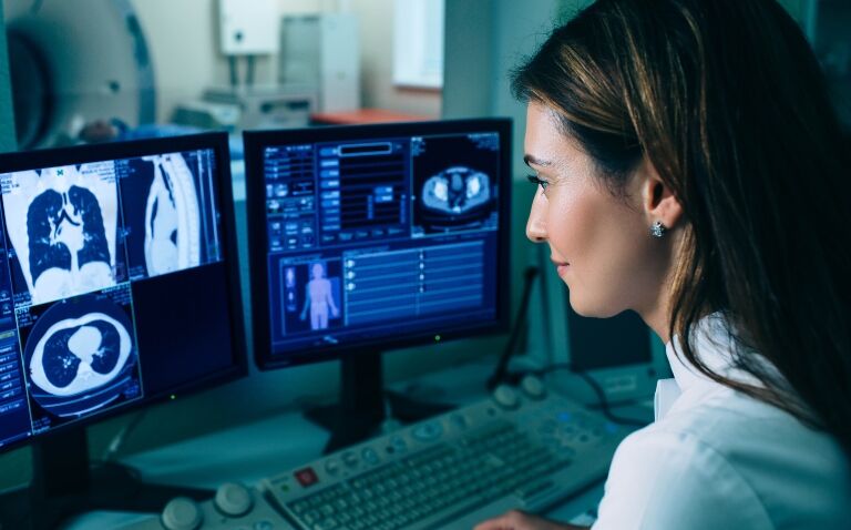
Speaking at Hospital Healthcare Europe’s Clinical Excellence in Respiratory Care event, our panel of three respiratory experts considered the use of diagnostic imaging for respiratory conditions. Dr Uta Hill, Jane Scullion and Dr Zaheer Mangera shared their views on how imaging techniques such as chest X-rays, CT scans and MRI can support the diagnosis and staging of respiratory diseases; the challenges that are being faced by the multidisciplinary team; and how new innovations are set to revolutionise patient care.
The diagnosis of respiratory conditions is reliant on a whole host of imaging techniques, holistic approaches and multidisciplinary input, all of which formed the basis of an in-depth discussion between three Clinical Excellence event panellists and chair John Dickinson, professor in sport and exercise sciences and head of the exercise respiratory clinic at the University of Kent.
Dr Uta Hill is a respiratory consultant at the Cambridge Centre for Lung Infection, part of the Royal Papworth Hospital NHS Foundation Trust. She is the clinical lead for cystic fibrosis and bronchiectasis co-clinical lead, contributing to the cystic fibrosis service and the hospital’s lung defence and immunology services.
Independent consultant respiratory nurse Jane Scullion spent many years working at Glenfield Hospital, part of the University Hospitals of Leicester NHS Trust, across the TB service, asthma clinics, COPD services and interstitial lung disease clinics. More recently, she has worked in medical negligence and long Covid clinics.
Dr Zaheer Mangera is a respiratory consultant and lung cancer lead at North Middlesex University Hospital NHS Trust who says he finds himself ‘knee deep in reviewing images’ every day as part of the image-heavy lung cancer pathway. He also works to support patients in quitting smoking and is involved in several tobacco dependency projects at a national level.
Together, the trio discuss how to successfully navigate imaging in diagnosing and staging respiratory diseases and the ways in which clinicians and the wider multidisciplinary team can support improved image interpretation, ultimate diagnostic capabilities and patient care.
How does imaging contribute to the way you diagnose respiratory disease?
Dr Hill: In my particular area, focusing on lung infection, we use CT scans a lot. That helps us to make the diagnosis of bronchiectasis, which is a structural lung disease predisposing to infections.
We then use CT scanning to assess the distribution of the bronchiectasis, the severity of the disease and the extent. This really helps us when we discuss it in our multidisciplinary meetings to plan how we treat these patients, how we manage them, where to target chest physiotherapy, which antibiotics or antifungals to use, and what other aetiologies might be necessary to assess further.
We are now able to use the low-radiation CT scans, particularly in my area of looking at lung infection. We can use that quite successfully to diagnose bronchiectasis. It does reduce the radiation exposure somewhat to the patients, which then may make it more acceptable.
Ms Scullion: As a nurse, I’m only allowed to do chest X-rays and CTs, although we do look at PET scans, MRI, and VQ scans. Working in interstitial lung disease is very multidisciplinary. We get a lot of chest X-ray abnormalities that are reported in, and we rely quite heavily on CT scans for diagnostics but also follow-up and prognosis.
Part of our therapeutic range of giving antifibrotics relied quite heavily on getting the right diagnosis for the right patient because, depending on your aetiology, you’ve got different treatments, so it was a lot more important. More recently, we have been allowed to give things for progressive lung disease – a lot of what we see is progressive.
We do pick up a lot of lung abnormalities that are actually just lung abnormalities that the patient gets really stressed and worried about but aren’t serious, and we will pick up some cancers and everything else.
Dr Hill: A few things that we have not mentioned, for instance, ultrasound. So, the value of ultrasound in looking at pleural effusions, evaluating them and then targeting areas for biopsy through thoracoscopy.
Often, it is also necessary to think about different types of CTs. So, we often use CTs with expiratory and inspiratory imaging to look for dynamic airway collapse, which can be a cause of recurrent coughing and chest infections but may not necessarily lead to bronchiectasis. But you would only pick it up on a CT scan if you asked for the inspiratory and expiratory imaging.
We use magnetic resonance imaging to longitudinally track changes in patients who are younger and where we want to limit radiation exposure. And that is also a key point: we are aware of the risk of radiation to the patients and the consequences that it can have, and that we keep thinking of modalities or ways of ensuring we minimise the risks.
How about the diagnosing and managing of lung cancer?
Dr Mangera: Thinking of the lung cancer pathway specifically, it usually starts off with a chest X-ray, usually requested in primary care or via the emergency department. An interesting question that we have to try to answer often is whether a patient should go directly to a CT scan of a chest and bypass the chest X-ray and that is becoming increasingly part of normal practice. Rather than delaying a patient’s diagnosis by wanting a chest X-ray first – that can be a straightforward process but sometimes it’s not – we are increasingly getting patients straight to CT scan where the index of suspicion for peripheral cancers is on the higher side and this is now reflected in national guidance as well.
It also reflects that fact that patients, as part of a lung cancer screening and targeted lung health checks, are going straight to CT scan so it doesn’t, make sense to introduce a chest X-ray step on a cancer pathway when that step doesn’t exist in a screening pathway.
In addition to the CT scan, that is very much the centre stone of everything that we do in the early part of the pathway, we also use nuclear medicine, PET-CT scan, which is an important test for staging someone’s scan.
For patients with lung cancer, we image pretty much every part of the body depending on the need. We will invariably use MRI when we are worried about disease of the brain or the spinal cord or bone disease, for example. We will employ ultrasound for image-guided biopsies, and so we tend to use a whole plethora of radiology techniques as part of the lung cancer pathway.
Is there a testing strategy or a hierarchy of scans ordered, or is it very dependent on what you are trying to investigate?
Dr Mangera: It’s very, very clear that there is a hierarchy of scans. If we think about the chest X-ray first, although it is a good test, there is lots of evidence about how you can pick up cancers even quite early on. The reality is that one in four lung cancers will have a normal chest X-ray – roughly speaking. Things will get missed, and there is a degree of human error with reporting.
I’ve seen what looks like quite obvious cancers being missed by very experienced radiographers. It does not happen very often, but we are looking at very subtle findings sometimes, and subtle findings can very easily trick you into looking normal, especially when you are sitting in a dark room looking at scans endlessly day after day. There is some scope for AI to assist reporting radiologists and radiographers. One way it might assist is it may focus the reporter’s attention into a particular area, for example. It’s not going to replace someone reporting, it’s not going to produce reports, but it will perhaps suggest that an area needs a closer look by the reporter.
Going onto CT scan, which is our bedrock. CT scan is fantastic at excluding cancer, it is not so good at confirming whether the abnormality that you are seeing is a lung cancer or not. I have seen all kinds of things that we thought would be a cancer coming back as not a cancer – typically infections. Even in the last month, I have told somebody they probably have a cancer, and it has come back as tuberculosis, for example.
More often than not, when you report a CT scan is showing cancer, particularly when there is a metastatic spread with mediastinal lymph node enlargement, for example, then it is very good at picking up lung cancers. But, every now and again, we find that we sometimes pick up diagnoses that are very good mimics of lung cancers.
And then, beyond a CT scan, the testing strategy is very much around what is going to get the staging of the cancer and PET-CT scan, invariably, is a tool for staging. It is uncommon that you need to go beyond a PET-CT scan to stage – except in the brain where an MRI scan will be superior to a PET scan in terms of staging.
What other techniques or diagnostic tools should be used alongside respiratory imagery to get a holistic picture of the patient and their diagnosis?
Dr Hill: I want to emphasise the importance of using radiology in conjunction with histology; microbiology, in my case; and cytology to hone down the diagnosis. In all these areas of respiratory medicine, the diagnosis and further management are made in a multidisciplinary team discussion, where consultants, nurses, physiotherapists and radiologists come together to make the diagnosis and plan the management.
Ms Scullion: It is probably around the history taking, because that tends to give you an awful lot. I only can speak from interstitial lung disease, but we get a lot of referrals in from rheumatology, we get a lot from cardiology as well. I think it’s really important that we don’t all look at our specific bit, that we look more generally at what is going on with patients.
So, their history, especially if there is going to be a drug reaction within the lungs, how long have they been taking it? What came first, was it the drug or was the progressive disease already there? For me, the patient is really central to it. That is why we are all working in multidisciplinary teams because we all bring a little difference to it. We have always benefited from having pharmacists in our team as well, because they’re very good at all these weird drug reactions that are actually causing lung abnormalities.
How do you make sure you get accurate diagnoses from imaging, and what challenges are currently being faced in this area?
Dr Mangera: A cornerstone of imaging is not the technique; it is the person reporting. If you do not have an experienced radiologist in thoracic radiology, then everything can look like a cancer, and everything will be reported as a cancer, or as interstitial lung disease, or an atypical infection, or whatever it may be. The recommendation will always be lung MDT.
This is an increasing problem, particularly in the UK setting, where there is a significant gap in the number of specialist radiologists, particularly thoracic radiologists. And with an ever-increasing market of outsourcing, where, in many Trusts, a large proportion of scans are reported outside of a Trust by private organisations. You may well get someone whose day job is a neuroradiologist, but at night they are doing thoracic radiology as part of trying to clear backlogs.
So, the thoracic radiology specialist within an MDT is key. But at the moment, I’m sure everyone is finding this, our MDTs are swamped with cases that are not being dismissed at the first round of reporting because of a lack of specialist thoracic radiologists reporting scans.
Dr Hill: As clinicians or nurses requesting imaging, we need to make sure we provide the relevant information. So, the smoking history, very brief details on whether they’re looking for infection and what the symptoms are, not just saying ‘coughing’ and as the only information. It really does help the radiographer or radiologists reporting if they do have all the relevant information on the request.
What other challenges do you currently face in respiratory imaging?
Dr Mangera: There are so many challenges for radiology because of the capacity issue of not having enough scanners and how far behind the UK is in terms of having physical infrastructure and the sheer number of scanners required for our population and the human resources required. We are far behind other countries like Germany and France, for example.
But in terms of day-to-day practice, I think incidental findings is the thing that’s choking our service more than anything. One thing we all have to remember is every time you perform a scan, you will find things that you are not looking for and are not relevant to the presenting complaint. What do you do with these things? And how do you know who has ownership of that problem?
With lung imaging, for example, we now get referrals from CT coronary angiograms and cardiac MRIs, we get referrals from our colorectal surgeons all the time because they do a CT chest everytime they do a CT colonogram for reasons that I still don’t fully understand. Patients coming in with any kind of complex trauma will have a CT head-to-toe – that’s the standard of care now. So, the sheer volume of incidental findings coming through the door has rocketed. While we have excellent national guidance on how to manage lung nodules, the sheer volume is causing a significant amount of pressure on the service.
That is before we even go into how that affects patients. Telling a patient that you have a lung abnormality, which is very unlikely to be a cancer but could be a cancer, depending on the psychology of that patient, they will take it well, or it will keep them up at night, potentially, for the next one or two years whilst they are under that surveillance period. I think incidental findings are a real problem.
Ms Scullion: An issue that we have is needing scanners that will take our larger patients. We’ve got a lot of patients who are over the weight limit for a lot of the investigations that we want to do, although you could argue, is it limited the information you get back just because of the population size?
Also, in secondary care, we can over-investigate people an awful lot because we are able to. I think it is about knowing when to do the investigations and also when to stop doing the investigations and always backing it up in terms of what’s happening with the patient, their symptoms and the good clinical history as well.
It’s also about learning to offer that reassurance to patients that what may have sounded alarming to them, actually isn’t. Because we check progression of disease, a lot of people who have maybe had TB in the past or sarcoid can worry about it being there, but it’s inactive and it’s just left a scar. So, scarring to patient may be very worrying, but to us it’s resolved.
What new technological advances might help improve respiratory diagnosis going forward?
Dr Mangera: If you speak to radiologists, AI is the big thing. The message is AI is not going to be the death of radiologists. The way AI work is it will help to support radiographers and radiologists in identifying problem areas. Specifically, AI can report dozens and dozens of scans within minutes and so what it will do is it will allow abnormal scans to be put to the front of the queue for a radiologist to report.
I think where the big value in AI will be is if a patient has a potential abnormality that warrants an earlier review, they’ll be pushed right to the front of the queue. That will make a huge difference in the current climate where, in some areas in the UK, patients and doctors are waiting weeks and weeks for the reports to come back. With the rollout of AI, hopefully we’ll be reassured that those with probable abnormal scans are going to have their scans reviewed much earlier on. That’s what I’m told about the added value of AI over the next five years.