This website is intended for healthcare professionals only.
Take a look at a selection of our recent media coverage:
27th June 2024
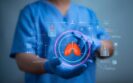
The opportunity to transform patient care using robotic techniques for lung transplantation is being championed and embraced at Vall d’Hebron University Hospital, spearheaded by Dr Albert Jauregui. Here, with an introduction from Helena Beer, Dr Jauregui discusses robotics within the context of complex lung surgeries, his increasing experience with this technique as well as the benefits and challenges he’s identifying and addressing, and the promising future of this enhanced surgery.
For many clinicians, the opportunity to support patients through a difficult time in their lives and have a profound and enduring impact on their health and wellbeing is a key reason for joining the profession, and transplantation is arguably one of the fields in which this can be achieved most acutely.
This was certainly among the determining factors in Dr Albert Jauregui’s career choice. ‘I decided to dedicate myself to thoracic surgery because it is a very attractive specialty within the field of surgery [as] you have many possibilities to help patients,’ he says.
As chief of the department of thoracic surgery and lung transplantation at the Vall d’Hebron University Hospital in Barcelona, Spain, Dr Jauregui is the driving force behind the adoption of medical technologies that have been making significant advances in the field so that his team can make an even bigger impact on clinical care and patient outcomes.
Robotically assisted, minimally invasive surgeries for lung cancer have been undertaken at the hospital for several years, but lung transplantation has still required aggressive surgery, which is something that Dr Jauregui is keen to change.
In 2023, his team performed Spain’s first two robotically assisted single-lung transplants, with both patients showing good improvement in the postoperative period and needing only mild pain medication after surgery – an outcome that the team is very happy with.
Bilateral lung transplants are next on the agenda, and Dr Jauregui and his team plan to perform five of these robotically assisted procedures this year and 10 in 2025 to build up the bank of evidence and continue working towards offering this type of innovative surgery to more patients.
Although the pool of lung donors has increased in recent years, thanks partly to relaxing the acceptance criteria and allowing new types of donors, the number still needs to be improved. The most important thing is to ensure a correct match between donor and recipient based on blood typing and immune characteristics to avoid organ rejection.
It is also essential to consider the graft size because large lungs can cause spatial problems in small thoracic cavities, and small grafts can be related to chronic dysfunction in the recipient. Therefore, it is crucial for lung transplant teams to try to find the best recipients for each donor.
Lung transplantation itself is considered one of the most complicated transplantation procedures due to the unique characteristics of the lungs – they are the only solid organs that have contact with the ‘outside world’ through breathing. Technically, it is a challenging surgery requiring collaboration with a large, experienced, multidisciplinary team to ensure the best results.
Lung transplant recipients are also particularly susceptible to infections and require high doses of immunosuppressive medications under strict control to avoid graft rejection and boost their immunity.
Thoracic robotic surgery has had a great impact on patients, especially in lung cancer where we see that its progress is unstoppable. The technical improvements offered by robotic surgery provide the patient with benefits in terms of recovery after surgery that are much better than traditional surgery.
Furthermore, the accompanying surgical precision makes the robotic technique extremely useful in lung cancer and transplantation and, thanks to the great experience of our transplantation team, we felt it had to be a logical evolution towards less aggressive and more precise surgeries.
Patients awaiting a lung transplant are usually significantly weakened and are traditionally offered a very aggressive open technique.
Our team performed robotically assisted surgeries with synthetic lungs in the lab before moving to large animal models. It was found that by deflating the lung and relying on the skin’s flexibility, we could use a smaller incision below the sternum to remove and insert the lungs.
Now, with the incorporation of robotic surgery for this type of patient, we have been able to reduce surgical aggression with preliminary results showing a substantial improvement in their recovery after a lung transplant.
The technique is still in its infancy and it’s at too early a stage to be able to operate on all the patients who are on the waiting list for a lung transplant using robotic surgery. Recipients tend to be a very heterogeneous patient group, but we are sure that in the very near future, it will become the surgical technique of choice.
As it is a surgery performed with small incisions and minimal invasion, the healing process will, of course, be quicker and less painful. This is especially beneficial because, as already said, these patients are usually frail due to the nature of their disease. Providing care in the least invasive way possible can only bring better results.
Robotic procedures account for 3% of all lung transplants performed at our centre. The technique is already defined, but greater patient numbers are needed to demonstrate its differences compared with traditional methods.
Our team will perform more robotic lung transplants each year, but it will take time to reach definitive conclusions. For now, the results regarding recovery and postoperative pain are encouraging. Patients who undergo robotic surgery need less analgesia and recover faster, but we still cannot reach absolute conclusions.
The most significant limitation is the widespread differences seen in terms of disease and degree of severity for patients on lung transplant waiting lists. As robotic lung transplantation is a very new technique, it requires performing a larger number of procedures and greater experience to generalise its use in more patients.
Another area for improvement is the surgeons’ experience with robotic surgery procedures themselves. Robotic surgery is increasingly being used, but it is still not widespread in general thoracic surgery programmes, and even less so in lung transplant programmes.
However, the worldwide interest in our findings in Barcelona makes the future promising, and we hope that more thoracic surgeons will join the movement.
This technology will undoubtedly improve in the future with the advent of more precise and even less invasive platforms. We are sure that organ transplants will become increasingly robotic in the future.
4th January 2024
With the results of the lung cancer screening SUMMIT study expected imminently, Helen Gilbert caught up with consultant respiratory physician Dr Neal Navani to discuss this research, promising new innovations in lung cancer diagnostics and what they might mean for the future of lung cancer care.
As Cancer Research UK’s Lung Cancer Centre of Excellence, University College London and University College London Hospital (UCLH) have been at the forefront of lung cancer innovations, pioneering diagnostic modalities such as endobronchial ultrasound.
This diagnostic focus is particularly pertinent as lung cancer is Europe’s biggest cancer killer, with 380,000 deaths across the continent in 2020 – a fifth of all cancer deaths.
In England, more than 60% of lung cancer patients are diagnosed at either stage three or four, and this late diagnosis is a frustration for Dr Neal Navani, lead consultant respiratory physician for lung cancer services at UCLH, as he says cure rates can be as high as 80-90% for patients whose small, early-stage lung cancer is detected.
Dr Navani, who is also the clinical lead of the UK National Lung Cancer Audit and clinical director for the Centre for Cancer Outcomes at the North Central London Cancer Alliance, has long been involved in pioneering research at UCLH to improve early detection and diagnosis.
And recent projects suggest there are further innovations on the horizon that have the potential to improve patient outcomes.
In May 2023, the largest lung cancer screening study of its kind in the UK drew to a close.
The four-and-a-half-year SUMMIT study was a collaboration between researchers from UCLH, University College London (UCL), the National Institute for Health Research, UCLH Biomedical Research Centre and GRAIL – a US healthcare company focused on the early detection of cancer.
Their aim was to identify lung cancer early among at-risk Londoners and support the development of a new blood test for the early detection of lung and multiple cancer types.
More than 13,000 people aged 55-77 from north and east London who had a significant smoking history were offered a blood test and a low-dose CT scan of their lungs. They were followed up at three months or immediately if a cause for concern was identified.
Dr Navani describes the research – results of which are expected imminently – as ‘a really fantastic, rich data set on which we can look to answer a lot of questions about detecting cancer early’.
He is particularly interested in developing a model that incorporates PET-CT scans to predict malignancy in screen-detected lung nodules. Often these appear like freckles on the lung, which may or may not be cancerous.
The challenge, he says, is working out whether they are malignant or benign, and currently this is done using a risk calculator developed in 2005.
It involves an injection of radioactive sugar before a PET-CT scan to see whether the nodule – or anything else for that matter – takes up the sugar. This then correlates with the risk of malignancy.
However, Dr Navani describes the current tool, which was developed in 2005, as out of date and prone to underestimating the risk of cancer in lung nodules.
Data from the SUMMIT trial are set to be used to develop and test a new risk calculator that takes into account more than 10 factors including family history, smoking and the size and appearance of nodules. It aims to accurately predict the chance of a nodule being cancerous.
‘We’re able to see whether sugar is taken up by that nodule in the lung – the idea being that small cancers use up more sugar than nodules that are not due to cancer,’ Dr Navani says.
‘Data for that work are being collected and developed. We’re pulling together data through other trials doing a similar thing and hopefully we’ll be able to clarify the role of PET-CT scanning for nodules in the next two years.’
The risk calculator will be compared against the existing model as well as others that do not include PET-CT scanning.
If found to be more accurate, the potential benefits are numerous and may include fewer patient investigations at lower cost, earlier treatment and reduced anxiety for those called in, Dr Navani explains.
UCL researchers are also using blood samples from the SUMMIT study to evaluate a blood test that can diagnose tumours earlier and detect 50 types of cancer, including lung cancer, with high accuracy.
Developed by GRAIL and an international team of researchers co-led by UCL, the test looks for tell-tale chemical changes to bits of genetic code – cell-free DNA – that leak from tumours into the bloodstream.
It was developed using artificial intelligence (AI) after researchers fed data on methylation patterns from the blood samples of thousands of cancer patients into a machine learning algorithm. It is said to identify many types of cancer, including bowel, ovarian and pancreatic, and can diagnose in which tissue the cancer originated with 96% accuracy.
But the potential of technology in bolstering cancer diagnosis doesn’t stop at AI. Another promising area of innovation is robotics.
Dr Navani is intrigued by the potential of this kind of diagnostic ability, and he is aware of robotic techniques that will be ‘the subject of research over the next year or two’.
He says: ‘We need to understand the cost effectiveness of robotic diagnosis of lung nodules. It’s potentially exciting.’
Earlier this year NHS clinicians at the Royal Brompton and St Bartholomew’s Hospital in London began a clinical study trialling a robotic-assisted bronchoscopy system.
Each hospital site is aiming to recruit around 50 patients with small lung nodules located in areas that are challenging to reach via traditional bronchoscopy.
The system combines software, robotic assistance and a flexible catheter with a camera to create a 3D roadmap of the lungs – much like a car’s sat-nav.
Doctors are directed to deep and hard-to-reach areas in each of the 18 segments of the lung, with the aim of removing tissue samples for biopsy with greater precision and accuracy.
The benefits of diagnosing a lung nodule accurately with a tiny camera could ‘open up a world of possibilities in terms of drug delivery, or ablation [to destroy cancerous nodules] in a controlled and accurate way,’ says Dr Navani. ‘I think in the next five to 10 years we’re going to see novel diagnosis and treatment options for our patients with early-stage lung cancer in particular.’
Another key development Dr Navani anticipates is the continued and increasing importance of collaboration, particularly when it comes to technology.
Endobronchial ultrasound (EBUS), one of the biggest innovations in respiratory medicine over the last 15 years, evolved from endoscopic ultrasound used in other clinical areas.
EBUS was trialled in the early 2000s by the UCLH research team, of which Dr Navani was a leading player, and uses a bronchoscope with a light, camera and integral ultrasound scanner to produce a detailed image inside the chest.
It enables doctors to take targeted needle biopsies of any enlarged lymph nodes and suspicious lesions while avoiding areas such as blood vessels.
Prior to this, patients at-risk of lung conditions required incisions to the chest under general anaesthetic, resulting in hospital stays and the possibility of complications or even death.
The arrival of EBUS in clinical practice in 2007 meant the diagnostic procedure could be performed on outpatients in under 30 minutes, with patients able to leave just one or two hours later.
‘It’s a very safe technique [and] in the last 10-15 years it’s really become a mainstay of diagnosis in respiratory medicine,’ Dr Navani acknowledges. ‘It started off very slowly but now in the UK there are 140 centres that are doing this technique and it’s been adopted globally for diagnosing lung conditions.’
Dr Navani believes the adaption of tools and techniques used in other clinical fields will continue to play a pivotal role in the advancement of lung cancer diagnostics and treatment. He points out, for example, that tumour ablation, which is used to treat lung and liver cancer, is now happening at a research stage for pancreatic cancer.
And this collaboration doesn’t just extend across clinical specialities. Imaging and information providers, including the likes of Fujifilm, also serve a vital purpose by providing increasingly innovative imaging solutions.
In June 2023, NHS England announced the national rollout of a targeted lung cancer screening programme to help detect cancer sooner and speed up diagnosis.
The rollout followed a successful pilot phase in which lung cancer scanning trucks carrying out on-the-spot chest scans operated from convenient locations such as football stadiums, supermarket car parks and town centres.
In September, NHS England announced that more than one million people had been invited for a lung cancer check via the scheme and almost 2,400 cancers had been caught – an impressive 76% of which were diagnosed at stage one or two.
‘That’s going to hopefully need innovative imaging solutions, particularly low-dose scanners, and I think we need to work with industry in terms of the use of artificial intelligence to help with the reporting of those scans,’ Dr Navani says.
Innovative diagnostic imaging techniques are certainly in development, and Dr Navani sees huge potential in new technologies for treating patients, too.
‘In terms of delivering novel therapies, in the future there may be a role for delivering drugs directly into the lungs, the pleural space or endobronchially, lymph nodes, or primary lung lesions,’ he says.
Dr Navani describes working in a hospital that is attached to a world-class university as ‘fantastic’ because it grants access to ‘extraordinary expertise’ spanning science, sociology, data science, computer science and engineering.
‘The research into lung cancer at UCL is really incredibly broad and, dare I say it, world leading, right the way through the basic science, biology and understanding how cancer develops and spreads and changes over time… to understanding the societal impact, equality and equity of care,’ he says.
According to Dr Navani, there appears to be a big difference in the outcomes of lung cancer patients based on socio-economic status.
‘We’ve really tried to address this in the National Cancer Audit, but it remains a significant challenge,’ he says. “A lot of this comes down to local resources… access to healthcare, equality and subsequent diagnosis and treatment in a timely fashion.’
Another major unmet need, Dr Navani says, is the 15% of patients with lung cancer who have never smoked and it’s here that ‘urgent research is needed’.
‘Given the high burden of lung cancer care, that’s a significant number of people – if you consider [non-smoking-related lung cancer] as a cancer in its own right it would be the seventh most common cause of cancer death,’ he says.
‘We’re really starting to get to grips with lung cancer in smokers but we are still at the early stages of understanding why people who’ve never smoked develop lung cancer. It would be important to predict who these people might be so that we can identify them at an earlier stage so hopefully their outcome will be better.’
The most pressing issue facing the NHS is limited resources, according to Dr Navani.
‘We simply don’t have enough scanners, radiologists, or space to do bronchoscopies,’ he states. ‘We’ve talked a lot about innovation but actually the most important thing that can be done to improve lung cancer care is for each hospital and primary care setting to have the appropriate resources to deliver what we know is already appropriate care, to drive out inequalities and drive everybody up to the best possible standards.’
While the future of funding for lung cancer care in the UK remains in flux, one thing is for certain: the research, expertise and drive to support the early diagnosis of patients remains, and Dr Navani’s commitment to supporting patients through innovative routes is stronger than ever.
9th November 2023
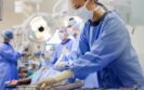
Specialist surgeons at Barts Heart Centre in London have performed a coronary artery bypass on a beating heart via robotic keyhole surgery for the first time.
Made possible using highly-sophisticated robot named the Da Vinci, the technology, which is funded by the Barts Charity, removes the need for open heart surgery. It therefore reduces the size of the would and subsequent recovery time.
The robot translates the surgeon’s hand movements in real time to four mechanical arms, which have a greater range of motion and precision. 3D images are beamed back to the surgeon via a high-definition screen.
A multidisciplinary team of surgeons, anaesthetists, nurses and perfusionists visited specialist centres across Europe to complete in-depth training before carrying out the heart surgery.
Commenting on the procedure, consultant cardiothoracic surgeon Dincer Aktuerk said: ‘Conventional coronary artery bypass graft (CABG) is still an excellent operation, however in selected patients a less invasive approach may be suitable. Where it would typically take patients several weeks to recover from a CABG, now, with robotic heart surgery of this kind, they are back to their old selves in days.
‘We are able to send patients home sooner, there is reduced pain and discomfort, faster recovery and much higher levels of patient satisfaction.
‘This is a major milestone for our hospital and for the UK as a whole. I would like to thank the entire team involved in this ground-breaking surgery.’
One of the first patients to undergo the heart surgery, 54-year-old Kemal said: ‘I was worried about the recovery time but two weeks after my operation, I am feeling good. I am now back on my feet and able to take short walks to the shop.
‘I want to give myself time before returning to work but I am pleased with my progress and feel very lucky to have had this kind of operation.’
The Barts Heart Centre at St Bartholomew’s Hospital is one of only two centres in the UK to offer robot-assisted heart surgery of this kind, and the only one in London. The robot at this hospital was the first in the UK to be dedicated to cardiothoracic cases.
26th July 2023
Surgeons in the UK lose an average of four working hours a week – equal to one working month a year – due to inefficient technology, according to a new survey by Censuswide on behalf of Medtronic.
The ‘State of Surgery in the UK’ survey explored surgeons’ attitudes towards the technologies they use in their role, the efficiency of them and the degree to which they enhance or hinder performance.
Some 79% of the 300 respondents said surgical care would be easier to deliver if technology was improved, and 58% agreed that technology in the operating room is inefficient and could impact the delivery of patient care.
In addition, 54% of surgeons reported spending time outside of hospital hours on administration that could be automated, and 56% of surgeons agreed time spent on administrative and logistical tasks could be reduced with better technology, which could free them up to focus on upskilling themselves and their team in other areas.
Commenting on the findings, Professor Sanjay Purkayastha, consultant upper GI and bariatric surgeon at Imperial College, NHS Healthcare Trust, and honorary professor at Brunel University, said: ‘The survey results reflect a challenge that many of us in the surgical community know all too well. For many surgeons, the lack of adequate technological support throughout the patient pathway leaves the surgical team perpetually short on time. Time that could be used on crucial analysis and training. An upgrade in the technologies available to surgeons is long overdue.
‘The enhanced efficiency and accuracy we gain from a more integrated and intelligent operating room are undeniable. In surgery, the benefits of being proactive, rather than reactive, are critical to maintaining a high quality of care. Digital technologies will be key to sustaining this. Unfortunately, these benefits remain out of reach for far too many in our field.’
Professor Naeem Soomro, consultant urological surgeon at Newcastle’s Freeman Hospital and Royal College of Surgeons Council member, added: ‘These findings validate and mirror our own research highlighting that the future of surgery lies in more forward-facing digital solutions. Robotics, data and artificial intelligence will allow the NHS to respond to current challenges around access, safety and sustainability of healthcare.’
This comes as the Royal College of Surgeons of England published a new guide covering some of the challenges – such as accessibility, variable outcomes and possible patient harm – and benefits of robotic surgery, including greater precision, freeing up hospital beds and improving patient recovery.
The guide, ‘Robotic assisted surgery: A pathway to the future’, also looks at the potential future application of robotics and makes recommendations to encourage sound governance practices that can lead to the safe adoption and expansion of robotic surgery in UK hospitals.
It proposes a structured pathway for established surgeons who want to transition to robotic-assisted surgery and identifies the relevant roles and responsibilities of key stakeholders for ensuring and maintaining safe autonomous practice in robotic surgery.
Nuha Yassin, consultant colorectal surgeon, robotics and minimally invasive surgery and RCS England Council lead for the future of surgery, robotics and digital surgery, said: ‘This timely new guidance will support the safe and structured introduction of robotic assisted surgery – and the fruitful collaboration between hospitals, surgeons and industry. It’s important for the surgical profession, led by RCS England, in collaboration with the surgical speciality associations, to take charge of all processes, accredit training centres and pathways and facilitate equity in access and training.
‘To benefit from the potential advantages, any investment in purchasing robots needs to be accompanied by proper planning for its introduction into the service with a focus on training, quality assurance and efficiency. This also needs to acknowledge the variable learning curve which can be long for some surgeons and theatre teams before these efficiencies can be observed at a large scale.’
IMPORTANT LINKS
MORE FROM COGORA
© Cogora 2025
Cogora Limited. 1 Giltspur Street, London EC1A 9DD
Registered in the United Kingdom. Reg. No. 2147432