This website is intended for healthcare professionals only.
Take a look at a selection of our recent media coverage:
7th July 2023
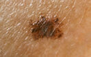
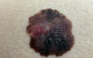
Cases of melanoma in situ are on the rise, but why is this the case and does it pose a greater risk of death among those diagnosed with the cancer? Clinical writer Rod Tucker considers the evidence.
Globally, there were an estimated 325,000 diagnoses of cutaneous melanoma – or simply melanoma – in 2020, with a corresponding 57,000 deaths. Based on current trends, it is estimated that there could be a 68% increase in melanoma-related deaths by 2040, although there are hopes that a melanoma vaccine used in the advanced stages of the disease might reduce these deaths.
Melanoma is now the 17th most common cancer worldwide, with cases steadily rising over the last 40 years. For example, the annual number of melanoma cases in men has increased from 837 per year between 1981 and 1985 to 6,963 per year between 2016 and 2018 – a more than eight-fold increase.
While invasive melanoma cases have increased substantially over the last 30 years, there has been a greater rise in the level of melanoma in situ, also known as a stage 0 melanoma, which are essentially benign lesions. The magnitude of the increase of melanoma in situ was revealed in a 2022 comparative study of invasive and in situ melanoma incidence in three predominately white populations in Australia, the US and Scotland.
The team considered the average annual percentage rate of change (AAPC) for the two types of melanoma and the results were somewhat surprising. While there was a significant increase in the AAPC for invasive melanoma in all three areas between 1982 and 2018, the largest increase was for melanoma in situ. For example, in white US males, the AAPC for invasive melanoma was 2.9 whereas for melanoma in situ, the figure was 8.7. A similar level of increase was seen across all three populations.
The purported culprit for the rise in melanoma cases is increased sun exposure, largely because of UV radiation. But does this really account for the observed increased incidence?
If greater exposure to sunlight believed to be responsible to the higher levels of melanoma, what does the research actually show?
In a meta-analysis of risk factors for melanoma focusing on sun – and therefore UV radiation – exposure, the authors extracted data from 57 studies and provided relative risk (RR) estimates for the different types of sun exposure. For total sun exposure, the risk of melanoma increased by 34% (RR = 1.34) and by 61% for intermittent exposure (RR = 1.61). In addition, there was a much stronger risk in those with a history of sunburn (RR = 2.03).
Yet, even in the worse case scenario, exposure to sunlight leads to no more than a two-fold increase in the risk of melanoma, much less than the actual eight-fold increased alluded to earlier. So what else could account for this discrepancy?
One possible reason for the higher incidence of melanoma in situ is that it is merely an artefact of increased skin cancer screening. This, it seems, is a more plausible explanation and there is some evidence to support it.
One study from 2012 suggested that today’s dermatopathologists are more inclined to diagnose melanoma than their predecessors. The study looked at biopsy specimens diagnosed as dysplastic nevi with severe atypia from 20 years earlier. Interestingly, researchers observed a general trend towards the reclassification of severely atypical melanocytic tumours as melanoma by all study participants. As the diagnosis of cutaneous melanocytic lesions relies on a pathologist’s visual assessment of biopsy material, it is ultimately somewhat subjective.
This point was clearly illustrated in a study from 2017 which considered the interpretation of pathologists’ diagnoses of melanocytic skin lesions. When a pathologist was asked to rate the same skin biopsies on two occasions, eight months apart, they demonstrated good reproducibility. However, the intraobserver reproducibility was much lower and, as the authors concluded, ‘diagnoses spanning moderately dysplastic nevi to early stage invasive melanoma were neither reproducible nor accurate.’
If the rise in overall melanoma diagnoses merely reflects an greater detection of melanoma in situ, is this still a cause for concern? To put it another way, to what extent do these benign lesions undergo malignant transformation and thus increase the risk of death?
This was the question addressed in a recent study published in JAMA Dermatology. The researchers examined the mortality risk among those diagnosed with a melanoma in situ. Following 137,872 patients with a first and only diagnosis of melanoma in situ, the researchers observed that 15 years after their diagnosis, the melanoma-specific survival was 98.4%.
Perhaps more interesting was the fact that the 15-year relative survival compared to the general population was 112.4%. In other words, virtually everyone diagnosed with a melanoma in situ was not only still alive 15 years later, but appeared to outlive those without the cancer.
But what if a melanoma in situ becomes invasive? Using a subgroup of 156,251 patients, the researchers observed that the 15-year cumulative incidence of a secondary primary invasive melanoma was 8.9% and 14.1% for a second primary melanoma in situ. Nonetheless, this had little impact on either melanoma or relative 15-year mortality which remained virtually unchanged at 98.2% and 126.7% respectively. Even among those who developed a second melanoma in situ, there was only a small reduction in both the 15-year melanoma and relative survival.
Based on these findings, questions have been raised as to whether it is time to stop skin cancer screening. In fact, this was the subject of a recent study by Australian researchers who tried to estimate the possible risk of over diagnosis of melanoma by the comparing the subsequent melanoma incidence and biopsy rates among people who either did or did not undergo skin screening.
When analysing all melanomas arising within the first year following recruitment, there was a 59% higher risk of a melanoma in situ but not for invasive melanomas. The authors calculated that the absolute risk of developing a melanoma after five years, in those who underwent screening would be 1.94% compared to 1.45% in those who were not screened. This, they said, equates to a number needed to screen to detect one excess melanoma of about 206.
Although this was a theoretical study, real-world data also suggests that failure to screen patients does not increase the risk of missed diagnoses. For example, the interruption of screening due to Covid-19 did not lead to unfavourable primary tumour characteristics of melanoma.
While it might seem prudent to continue screening but only for high risk patients, this is by no means an easy task given how the risk factors for melanoma are multi-factorial and based on both genetic and personal attributes.
With the development of new AI-based technology, it is anticipated that skin cancer screening could become both faster and possibly more targeted. Nevertheless, medico-legal concerns of a missed diagnosis are unlikely to recede in the near future, prompting an increased level of screening.
Perhaps it is time to raise the diagnostic bar for biopsies and avoid the labelling of benign lesions like melanoma in situ, especially given the devastating impact of a cancer diagnosis – even if unwarranted – on a patient’s wellbeing.
1st February 2023
In a study published in Science Advances, researchers from the Institute of Cancer research in London, have identified how cancer cells can either shrink or become super sized as a means of survival, in the face of drug treatment or any other challenges that might occur within their environment.
Although it is acknowledged that there are differences in the size of cell types, there is also a recognition that for proliferating cell types, there is little variation in cell size. This implies that there is some mechanism or checkpoint, responsible for ensuring that cells maintain a particular size during the proliferative phase. Clearly, this checkpoint is disturbed in cancer and it is known that cancers will arise due to the accumulation of mutations in genes and which alter the normal proliferation, differentiation and death process.
In the current study, researchers used a combination of biochemical profiling and mathematical analyses, to examine how genetic changes affect the size of cancer cells. They focused on skin melanoma cells which are known to be driven by two genetic mutations, a BRAF gene (60%) and an NRAS mutation (20 – 30%) and examined differences in the size and shape of these cells with the two mutations.
Interestingly, the team found that BRAF-mutant cancer cells were very small, whereas NRAS-mutant cells were much bigger and larger still, when the cells were drug resistant. It seemed that small cells were able to better tolerate DNA damage, due to high concentrations of DNA repair proteins such as PARP, BRCA1 or ATM1.
The researchers believed that their findings might help clinicians to decide upon the choice of treatment. For instance, small cells were likely be more vulnerable to PARP inhibitors, i.e., drugs affecting the proteins responsible for DNA repair. In contrast, the larger NRAS-mutant cells, already had lots of DNA damage, which the cells were unable to repair and hence unlikely to respond well to PARP inhibitors, whereas immunotherapy might be a better option. The team think that both the BRAF and NRAS mutations may cause changes in cell size by affecting levels of a protein known as CCND1, which is involved in cell division, growth and maintenance of the cytoskeleton, as well as its interactions with other proteins.
Commenting on the study, lead author, Professor Chris Bakal, from the Institute of Cancer research, said ‘Cancer cells can shrink or grow to enhance their ability to repair or contain DNA damage, and that in turn can make them resistant to certain treatments.’ He added that the study had a potential diagnostic value. For instance, ‘by looking at cell size, pathologists could predict whether a drug will work, or if the cells will be resistant. In the future, it might even be possible to use AI to help guide the pathologist, by making a rapid assessment about the size of cells and so the treatments that are most likely to work.’
29th December 2021
Having pre-existing heart disease (PEHD) seems to improve the chance of survival to hospital after experiencing an out-of-hospital cardiac arrest. This was the somewhat counter-intuitive conclusion of a retrospective analysis by a team from the Department of Cardiology, Amsterdam UMC, The Netherlands.
Out-of-hospital (OOH) cardiac arrests are a common problem and in one European study of 37,054 such arrests, the hospital survival rate was only 26.4%. Patient characteristics are undoubtedly a potentially important contributor to overall survival and in particular, the presence of existing heart disease, although the impact of this factor on survival has been poorly studied. In one such study, having ischaemic heart disease led to a 50% improved odds of survival. However and in contrast, a second study showed that PEHD was an independent predictor of a poorer survival after an OOH cardiac arrest.
Given these contradictory findings, the Dutch researchers set out to explore the relationship between OOH cardiac arrest and prior cardiovascular disease. They retrospectively examined data from the AmsteRdam REsuscitation STudies (ARREST) registry, which contains information on all OOH cardiac arrests in the Northern part of Holland. Patient’s medical histories were obtained from their GP’s medical records for evidence of a wide range of cardiac diseases and patients were dichotomised as either having or not having PEHD. For the purposes of the study, the primary outcomes were survival to hospital admission and survival to hospital discharge and this data was obtained from hospital records. Secondary outcomes were a shockable initial rhythm (SIR) and acute myocardial infarction (AMI) as an immediate cause of OOH cardiac arrest. Using regression analysis, the team examined the association between PEHD and survival to hospital and survival to discharge and adjusted models for several co-morbidities.
Findings
A total of 3760 OOH cardiac arrest patients with a mean age of 68 years (70.9% male) were included in the analysis. Overall, 48.1% of the cohort had PEHD and on average were slightly older (mean age 71.4 vs 64.7, p < 0.001) and with a higher incidence of cardiovascular risk factors e.g., hypertension (59.3% vs 42.2, p < 0.01), obesity and hypercholesterolaemia.
Among those with PEHD, 41.9% survived to hospital admission compared to 38% of those without prior cardiovascular disease (p = 0.014). The presence of existing heart disease was associated with an increased survival odds after adjustment for all covariates (adjusted odds ratio, aOR = 1.25, 95% CI 1.05 – 1.47). However, prior cardiac disease was not associated with survival to discharge in fully adjusted models (aOR = 1.16, 95% CI 0.95 – 1.42).
Among 1680 patients with SIR, prior heart disease was also not associated with survival to hospital (aOR = 1.12, 95% CI 0.90 – 1.39) but prior heart disease was associated with a lower proportion of AMI (aOR = 0.33, 95% CI 0.25 – 0.42).
In their discussion, the authors recognised that their findings were somewhat counter-intuitive and were unable to explain the result. Nevertheless, they concluded that survival gains after an OOH cardiac arrest applied to a wide range of patients with prior heart disease.
Citation
van Dongen LH et al. Higher chances of survival to hospital admission after out-of-hospital cardiac arrest in patients with previously diagnosed heart disease Open Heart 2021
7th October 2021
Community-acquired pneumonia (CAP) is an infection acquired in the community, i.e., outside of a hospital setting. The worldwide incidence of community-acquired pneumonia has been estimated to vary between 1.5 to 14 cases per 1000 person-years. Mortality rates for CAP are very low (< 2%) for patients treated in the community but increase among those hospitalised (5 – 20%) and are higher still (up to 50%) for patients who are admitted to intensive care. Treatment of CAP involves the use of empirical antibiotics and several guidelines exist for the management of CAP. Moreover, evidence suggests that guideline-concordant prescribing for CAP is associated with improved health outcomes and lower resource use in adults. But to what extent would guideline discordant antibiotic prescribing impact on health outcomes and mortality?
This was the question posed by a team from the Department of Emergency Medicine, Seoul National University Bundang Hospital, Republic of Korea. The team undertook a retrospective analysis of adult patients with severe CAP, hospitalised in the emergency department (ED) after the diagnosis of severe CAP, defined by the 2007, Infectious Diseases Society of America/American Thoracic Society guidelines. For the treatment of severe CAP, the guidelines recommend a beta-lactam antibiotic plus either a macrolide or fluoroquinolone. Among penicillin allergic patients, a respiratory fluoroquinolone plus aztreonam is recommended. Data on prescribing , together with demographic and co-morbidity information were obtained from hospital medical records. Patients were then categorised as either being prescribed guideline concordant antibiotics or guideline discordant antibiotics. Propensity score matching was used to reduce selection bias and 30-day survival was estimated with logistic regression.
Findings
A total of 630 patients were included, of whom 179 (28.4%) died within 30 days of being hospitalised. After propensity matching, a total of 255 individuals were included in each group with an approximate age of 75 years (66% male). After propensity matching, guideline discordant prescribing was significantly associated with 30-day mortality (hazard ratio, HR = 1.43, 95% CI 1.05 – 1.93, p = 0.022). In addition, 30-day mortality was found to be lower in the guideline concordant group (23.9% vs 33.3%, concordant vs discordant, p = 0.024).
Commenting on these findings, the authors noted that 43% of patients were prescribed guideline discordant antibiotics for severe CAP and concluded that this was independently associated with 30-day survival.
Citation
Hyun KS et al. Antibiotic prescription consistent with guidelines in emergency department is associated with 30-day survival in severe community-acquired pneumonia. BMC Emerg Med 2021.
12th August 2021
Among critically ill patients, intravenous fluids (IV) are used for intravascular volume replacement. Administration of such fluids is extremely common and it has been estimated that every day, over 20% of patients within an intensive care setting receive fluid therapy. In general terms, fluid therapy is required for several indications including impaired tissue perfusion, low cardiac output and abnormal vital signs, e.g., blood pressure, heart rate or urine output. The most commonly used IV fluid is saline solution (0.9% sodium chloride) although in recent years, there has been emerging evidence that IV fluids other than saline in critically ill patients may have a more favourable impact on mortality. Balanced IV fluids, for example, have been designed to be more aligned with the composition of serum and may have some advantages over saline. For example, one study in patients with sepsis, concluded that resuscitation with balanced fluids was associated with a lower risk of in-hospital mortality. In a 2018 study among critically ill adults, the use of a balanced crystalloid rather than saline, produced a lower rate of death compared to saline. The use of a balanced solution rather than saline has several other potential advantages, particularly in relations to adverse effects, since saline contains a higher concentration of chloride ions and has been associated with a hyperchloraemic metabolic acidosis and acute kidney injury.
However, the overall benefit of using a balanced IV fluid rather than saline is not always superior. For instance, the use of a balanced crystalloid did not reduce the incidence of acute kidney injury compared to saline within an intensive care unit (ICU). In trying to provide much needed clarity, the Balanced Solution versus Saline in Intensive Care Study (BaSICS) by a Brazilian group of clinicians was undertaken to compare the effectiveness and safety of balanced crystalloids compared with saline in critically ill patients. This trial undertaken at 75 intensive care units in Brazil, randomised patients admitted to an ICU to either saline or a balanced solution and the primary outcome was 90-day survival.
Findings
A total of 10,520 critically ill patients with a mean age of 61.1 years (44.2% female), were randomised to either balanced fluids or saline and patients in both groups received a median of 1.5 litres of fluid during the first day of enrolment. Of the whole cohort, 60.6% of patients had hypotension or vasopressor use and 44.3% required mechanical ventilation at enrolment. Within 90 days of enrolment, 26.4% of those assigned to balance fluids died compared to 27.2% given saline (adjusted hazard ratio, aHR = 0.97, 95% CI 0.90 – 1.05, p = 0.47).
The authors concluded that despite the potential advantages of balanced crystalloids over saline, there were no apparent mortality benefits.
Citation
Zampieri F et al. Effect of Intravenous Fluid Treatment with a Balanced Solution vs 0.9% Saline Solution on Mortality in Critically Ill Patients. The BaSICS Randomised Clinical Trial. JAMA 2021
9th July 2021
Breast cancer is the most common cancer in women worldwide and in 2020, there were 2.2 million cases and nearly 685,000 deaths. Breast cancer is a heterogenous disease with various types and different sensitivities to treatment, e.g., oestrogen receptor (ER) positive, progesterone receptor (PR) positive and hormone receptor (HR) negative. Among those with ER positive tumours, treatment with adjunctive therapy such as tamoxifen for five years is recommended. Nevertheless, even after treatment with tamoxifen, breast cancer can return over the intervening years, metastasise and lead to death. The use of clinical breast cancer markers such as tumour size or grade can be used to provide estimates of survival for up to 10 years and both increased tumour size and grade are associated with a reduced short-term survival. However, what is far less clear, is whether any of these markers are associated with longer term survival.
Given that both ER-positive and ERBB2-negative (i.e., HER2) disease are associated with a continuous risk of recurrent disease after the primary diagnosis, a team from the Department of Oncology and Pathology, Karolinska Institute, Stockholm, Sweden, sought to determine whether both clinically used markers and treatment with tamoxifen were associated with long-term survival in patients with breast cancer. The team turned to data from the Stockholm tamoxifen (STO-3) trial, which enrolled postmenopausal women with lymph-node negative breast cancer and tumours less than 30mm in diameter, between 1976 and 1990 and who were randomised to tamoxifen 40mg daily or no endocrine therapy. In 1983, women with no recurrence after 2 years on tamoxifen, were given the drug for a further 3 years. The team undertook an analysis of tissue samples that were available from patients in the study. The tumour grade (1 to 3) had been assessed in 2014 by a pathologist and tumour size was categorised as T1a/b if 10mm or less, T1c if 11–20mm and T2 if larger than 20mm. All patients had a unique national registration number and which was linked with a cancer registry, allowing the team to obtain long-term follow-up data after 25 years.
Findings
The sample contained 565 postmenopausal women with a mean age of 62 years. Just over half (52.2%) of tumours were graded as T1c and nearly a third (30%), T1a/b. Patients with T1a/b tumours had the best long-term survival (88%) compared to those with T1c tumours (76%) or T2 tumours (63%). Similarly, the highest level of survival (81%) occurred in those with grade 1 tumours compared with grade 3 tumours (65%). Using this data, the team estimated a 69% reduced risk of disease recurrence for those with T1a/b sized tumours (hazard ratio, HR = 0.31, 95% CI 0.17–55). In terms of treatment, those given tamoxifen with grade 1–2 tumours also experienced a significantly reduced risk of disease recurrence (HR = 0.24, 95% CI 0.07 – 0.82) compared with those who did receive the drug. However, use of tamoxifen also produced a significant survival benefit in those with T2 tumours (HR = 0.34).
The authors concluded that tumour size, followed by grade and use of tamoxifen were independently associated with long-term survival in breast cancer.
Citation
Dar H et al. Assessment of 25-Year Survival of Women with Oestrogen Receptor–Positive/ERBB2-Negative Breast Cancer Treated with and Without Tamoxifen Therapy. A Secondary Analysis of Data from the Stockholm Tamoxifen Randomised Clinical Trial. JAMA Netw Open 2021.
9th October 2020
It is thought that skin transmission is just one of many potential routes although little is known about how long the virus can survive on the surface of not only skin or other surfaces. In a new study by a team from the Department of Infectious Diseases, Kyoto University, Japan, researchers used skin samples obtained from forensic autopsy samples, collected one day after death and found that the virus can survive for several hours but that it is quickly eliminated after washing with a disinfectant. The team used influenza virus (IV) for comparative purposes but also examined the survival of both viruses on several difference surface materials such as stainless steel, glass and polystyrene. The survival of the viral samples on these different surfaces were also examined after being mixed with mucus samples. In all cases, the samples were incubated at 25oC for 30 minutes and the content of both influenza and COVID-19 analysed.
Findings
The results showed that COVID-19 survived for considerably longer than IV on all tested surfaces. For example, IV survived for up to 11 hours on stainless steel compared to 85 hours for COVID-19. The virus also survived for a similar time on glass but for only 58 hours on polystyrene. In contrast, COVID-19 managed to survive for only 25 hours on stainless steel compared and for nearly 24 hours on glass when mixed with mucus. When directly in contact with human skin, COVID-19 survived for 9 hours compared to 1.82 hours for IV but this was reduced to only 4 hours when in mucus. However, mixing with 80% ethanol reduced COVID-19 survival time in mucus to less than 15 seconds.
Commenting on their findings, the authors noted that because COVID-19 can survive for up to 9 hours on human skin it poses a significant risk for transmission but the study also highlighted the importance of hand washing with ethanol which inactivated both COVID-19 and IV viral particles within less than 20 seconds.
Reference
Hirose R et al. Survival of SARS-CoV-2 and Influenza virus on the human skin: importance of hand hygiene in COVID-19. Clin Infect Dis 2020; https://doi.org/10.1093/cid/ciaa1517
IMPORTANT LINKS
MORE FROM COGORA
© Cogora 2025
Cogora Limited. 1 Giltspur Street, London EC1A 9DD
Registered in the United Kingdom. Reg. No. 2147432