This website is intended for healthcare professionals only.
Take a look at a selection of our recent media coverage:
6th February 2024
There will be more than 327,600 premature deaths across Europe if there is a delay in fully aligning the EU’s Ambient Air Quality Directive (AAQD) with World Health Organization (WHO) Air Quality Guidelines (AQG) 2021, according to a new report.
Writing in the International Journal of Public Health, doctors and experts from European Respiratory Society (ERS) and other health groups called for immediate action on European air pollution to ensure alignment with the AQG by 2030.
They warned that not doing so will result in poorer health for millions of people and widen the health inequality gap between western and eastern Europe.
Research from Europe indicates a strong link between air pollution and millions of new cases of asthma, COPD, acute respiratory infections, lung cancer and other serious illnesses each year.
The report stated: ‘This is a historic opportunity towards clean air in Europe for all that could prevent hundreds of thousands of premature deaths and millions of new cases of non-communicable diseases every year, as well as improve health of all European citizens.
‘Full alignment with WHO AQG would enhance children health in Europe by improving lung function and reducing asthma and respiratory infections burden. Achieving the WHO AQG would also reduce healthcare costs, social, environmental and health inequalities, boost economic growth, and help mitigate the adverse effects of climate change.’
This urgent call for action against air pollution comes as trilogue discussions between the EU Commission, Parliament and Council continue, with a deal to be reached soon regarding revisions to the EU’s AAQD.
The current proposals, the report said, leave out full alignment with WHO AQGs and allow delays in achieving air pollution limit values up to the 2040 for countries whose gross domestic product per capita is below EU average.
The report highlighted that ‘using poverty as an excuse to fail to act is the opposite of what countries in Europe need’.
It concludes: ‘We strongly urge the EU environment ministers to put European health and environmental justice at the core of their political aspirations.
‘This is a unique public health opportunity for EU Member States to follow the scientific evidence and listen to the concerns of citizens.’
Commenting on the report, Professor Zorana J. Andersen, ERS Environment and Health Committee chair, said: ‘Allowing additional delays in reaching new EU air quality standards, differentiated based on GDP, is completely unacceptable to the ERS community. A delay would widen existing inequalities in air pollution levels and health burden between east and west.
‘Children and adults in eastern European countries have already been breathing the most polluted air in Europe and suffering from related lung diseases for far too long. We need fair, ambitious new EU air quality legislation that values the health of all Europeans equally.
‘A new Air Quality Directive must provide clear vision and support to speed up, and not delay, much needed air pollution reductions in eastern Europe, in order to improve health and wellbeing, and achieve clean air for all in Europe, as soon as possible.’
In September 2023, the ERS issued a consensus statement on climate change and respiratory health, including guidance on how global warming can be addressed in clinical practice.
And in November 2023, the WHO published a new operational framework for building climate-resilient, low-carbon and sustainable health systems and address climate-related health risks to safeguard health.
11th May 2023
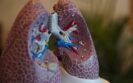
Fergus Gleeson has long held an interest in MRI scanning techniques that use hyperpolarised xenon – an odourless gas – to detect early-stage lung disease. Here, he discusses his interest in xenon, how the gas works in practice and his latest research findings in relation to long-Covid.
In 2020, Fergus Gleeson, professor of radiology at Oxford University and consultant radiologist at Oxford University Hospitals NHS Foundation Trust, turned his attention to a small study of post-Covid patients. They were experiencing breathlessness several months after being discharged from hospital, despite CT scans that appeared to show their lungs were normal.
The findings from this small study prompted Professor Gleeson to widen the research to investigate possible lung damage in non-hospitalised, post-Covid patients who continue to experience breathlessness many months after infection.
He is the principal investigator of the EXPLAIN study, one of 19 projects to receive nearly £40m from the National Institute for Health Research to improve understanding of long-Covid, from diagnosis and treatment through to rehabilitation and recovery.
I’ve worked in the xenon field since 2007/08 to identify patients with obstructive lung disease and interstitial lung disease, as have other researchers, including Professor Jim Wild, who is the head of imaging and NIHR research professor of magnetic resonance at the University of Sheffield. [Indeed, Wild pioneered the method, development and applications of hyperpolarised xenon MRI used in the EXPLAIN study.]
Xenon is unique in its ability to measure gas transfer in the lungs. The MRI scanner is programmed to detect signal from polarised xenon. The oxygen in the normal air we breathe isn’t used because it does not give enough MRI signal. Xenon behaves just like oxygen, so radiologists can use it to observe how it moves from the lungs into the blood stream and highlight any areas where it is not flowing well between the airways, gas exchange membranes and capillaries in the lungs.
The patient lies in an MRI scanner on a magnet with a vest like coil around their chest and breathes in one litre of inert gas xenon through a plastic straw. It is possible to watch the physiology of a patient’s lungs in real time and see where the xenon might be held up, how fast it moves, whether it gets to the alveoli and from this assess how well the exchange of oxygen and carbon dioxide happens in the blood. The degree of gas exchange is colour coded with areas of more damaged lungs being darker or a different colour to areas with normal exchange.
The scan times may be very short, for example a single sequence may only require the patient to breathe in and hold their breath for 14-20 seconds. There’s no radiation exposure so it can be repeated over time to see whether the changes to the lungs improve. If the research proves to be clinically useful, the equipment could be set up in multiple regional centres in a relatively short space of time. All that is needed is a cylinder of xenon and the polariser – which is about the size of a large chest of drawers. The MRI scanner would also need to be modified to detect the signal from the xenon gas.
Ours is located in the radiology department at the Churchill Hospital in Oxford. There’s one in Sheffield University’s imaging department, one is being installed at University College Hospital London and one at Manchester University. There’s also a couple in Denmark, two in Germany, several in the United States and at least one in Canada.
Some people were complaining of persistent breathlessness and fatigue for many months after the Covid-19 infection, so we conducted a small study on patients aged between 19 and 69. None of them required intensive care or ventilation, and standard CT scans showed no significant lung damage. The hyperpolarised xenon MRI scans revealed signs of lung damage for a number of the patients who were reporting breathlessness.
We then broadened the study out to include non-hospitalised individuals who had tested positive for Covid-19 across a range of age groups. Now, there are many different arms of the study and we’re working with three other hospital sites: Sheffield, Manchester and Cardiff. Hundreds of thousands of people in the UK continue to experience symptoms months after having Covid-19, with breathlessness one of the most commonly reported symptoms.
We hope to identify what’s causing patients with long-Covid breathlessness and essentially give them answers as to how long they might have these abnormalities and whether they’ll get better.
We’re using hyperpolarised xenon MRI scans to investigate possible lung damage in long-Covid patients who have not been hospitalised with Covid-19 but who continue to experience breathlessness. The full study will recruit around 400 participants following on from our two initial pilot studies. The EXPLAIN study will include:
We detected significantly impaired gas transfer from the lungs to the bloodstream in long-Covid patients when other tests were normal. These patients had never been in hospital, did not have an acute severe illness when they had their Covid-19 infection, and some had experienced symptoms for a year after contracting it. The important questions that now need answering are how many patients with long-Covid will have abnormal scans, what the significance of the abnormality we’ve detected is, what the cause of the abnormality is and what its longer-term consequences will be. Once we understand the mechanisms driving these symptoms, we will be better placed to develop more effective treatments.
We need to complete recruitment and analyse the data, so we have some way to go before fully understanding the nature of the lung impairment that follows Covid-19 infection. However, the clinical-academic collaboration in EXPLAIN and other studies funded by NIHR are fundamental steps towards understanding the biological basis of long-Covid. This, in turn, will help us to develop more effective therapies.
4th May 2023
Type 2 diabetes has been shown to cause lung disorders for the first time in a new study funded by Diabetes UK.
In the largest-ever genetic study to explore how genes affect blood sugar levels and health outcomes, researchers from Imperial College London concluded that lung disorders should now be considered a complication of type 2 diabetes.
When examining the impact of blood sugar levels on lung function, the researchers found that people with type 2 diabetes who had a three-fold increase in average blood sugar levels, experienced a 20% drop in lung capacity and function.
The findings, presented at the recent Diabetes UK Professional Conference 2023, highlight the need for healthcare professionals to be alert to lung complications within patients with type 2 diabetes, alongside kidney disease, heart attack and strokes.
More than five million people in the UK live with diabetes, and 90% have type 2. These patients often have dangerously high blood sugar levels caused by the body either not making enough insulin or not responding to the insulin that is made.
Chronically high blood sugar levels can damage organs and tissues, causing kidney failure, eye and foot problems, heart attacks and strokes. Previous research has shown that lung conditions, including restrictive lung disease, fibrosis and pneumonia, are more common in people with type 2 diabetes, but no causal link had been established.
Using statistical techniques, the researchers analysed data from almost 500,000 participants on 17 major studies, including the UK BioBank, to determine whether there was a causal link between impaired lung function and high blood sugar levels. Lung function was measured using two standard spirometry tests used to diagnose lung conditions.
High blood sugar levels in people with type 2 diabetes were shown to impair lung function directly. Statistical modelling of the study data showed that an increase in average blood sugar levels from 4 mmol/L to 12 mmol/L could result in a 20% drop in lung capacity and function.
Dr Elizabeth Robertson, director of research at Diabetes UK, said: ‘These results are a reminder of the seriousness of type 2 diabetes and the importance of supporting people with the condition to manage their blood sugar levels so they can live well with the condition and avoid future complications.
‘Lung conditions can be life-changing and life-limiting, and it is crucial that healthcare professionals are aware of the impact of high blood sugar levels on lung health.’
This news story was originally published by our sister publication Nursing in Practice.
4th July 2022
Cannabis use is not associated with more respiratory-related visits to an emergency department in comparison to those who do not use the drug although it is associated with a greater proportion of overall emergency department visits.
This was the main conclusion of a propensity-matched study by a group of researchers from Ontario, Canada.
Cannabis (or marijuana) is the most commonly used addictive drug after tobacco and alcohol. The use of cannabis is associated with respiratory problems such as chronic bronchitis symptoms and large airway inflammation and in fact, heavy use may lead to airflow obstruction.
Despite this evidence of adverse respiratory effects, a 2018 systematic review concluded that there was low-strength evidence that smoking cannabis was associated with cough, sputum production, and wheezing and that there was insufficient evidence of an association between use of the drug and obstructive lung disease.
Nevertheless, one study has suggested that daily cannabis smoking, even in the absence of tobacco, is associated with an elevated risk of health care use for various health problems.
With some uncertainty over the respiratory effects of cannabis, in the present study, the Canadian team wanted to examine the magnitude of the association between the use of cannabis and adverse respiratory-related emergency department visits.
They conducted a retrospective analysis linking health survey and health administrative data for residents of Ontario. Individuals who self-reported any use of cannabis (the exposed group) within the past year were matched 1:3 (to increase the sample size) with control individuals, which were those who self-reported no use of the drug.
The primary outcome for the study was a respiratory-related emergency department visit or hospitalisation which included both upper and lower respiratory tract infections, respiratory failure, asthma or COPD as the reason for presentation at the hospital. As a secondary outcome, the team assessed all-cause emergency department visits.
Cannabis use and respiratory-related hospital visits
A total of 35,114 individuals were included in the analysis, of whom, 6,425 with a mean age of 32.2 years (38.8% female) were self-reported cannabis users. Overall, 42.5% of those using the drug did so less than once a month with a much smaller proportion (10.5%) reporting daily use.
The odds of a respiratory-related emergency department visit or hospitalisation was not significantly different between the exposed and control group (odds ratio, OR = 0.91, 95% CI 0.77 – 1.09, p = 0.32). Despite this, there was a greater odds of all-cause emergency department visits among those who used cannabis (OR = 1.22, 95% CI 1.13 – 1.31, p < 0.0001).
The most frequent reason for the emergency visits among those using cannabis was for acute trauma (15.1%) although interestingly, this was followed by respiratory problems (14.2%).
The authors concluded that while there were no differences in the proportion of respiratory-related hospital visits between the two groups, all-cause hospital visits were significantly higher among those who. self-reported cannabis use.
They added that based on these findings, recreational use of the drug should be discouraged.
Citation
Vozoris NT et al. Cannabis use and risks of respiratory and all-cause morbidity and mortality: a population-based, data-linkage, cohort study BMJ Open Respir Res 2022
IMPORTANT LINKS
MORE FROM COGORA
© Cogora 2025
Cogora Limited. 1 Giltspur Street, London EC1A 9DD
Registered in the United Kingdom. Reg. No. 2147432