This website is intended for healthcare professionals only.
Take a look at a selection of our recent media coverage:
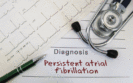
18th November 2022
A model combining features of an ECG and radiomics data derived from cardiovascular magnetic resonance imaging improved the detection of atrial fibrillation (AF) in women to a greater extent than either model alone according to the findings of a study by Spanish and UK researchers.
Atrial fibrillation is the most common cardiac arrhythmia, characterised by an irregular heart rhythm and an often abnormally rapid heart rate and globally has been estimated to affect 0.51% of the population. AF is diagnosed from an ECG in which typically, P-waves are absent and there is both a chaotic baseline and an irregular ventricular rate. Cardiac magnetic resonance (CMR) is the reference imaging modality for assessment of cardiac structure and function and CMR radiomics has the potential to improve diagnostic accuracy. Moreover, Cardiac MRI of the atrial substrate is not only a tool for management and treatment of arrhythmia, but also to individualise the prevention of stroke and major cardiovascular events. But what remains unclear is whether the use of a CMR-based radiomics model can identify patients with AF and more importantly, if addition of a model that uses ECG-derived data, would further enhance the potential to detect AF.
In the present study, researchers examined the feasibility of combining a CMR-derived radiomics model and one based on ECG data. The team used information from the UK Biobank and identified patients who had both an ECG and CMR scan and compared their findings with healthy controls. Models were assessed using the area under the receiver operating characteristics curve (AUC) and associated sensitivity and specificity.
ECG and radiomics model and atrial fibrillation detection
A total of 32,121 participants with a mean age of 63 years (51% female) were included and of whom, 495 (63% male) had AF.
Overall, the AUC for the combined model was similar to the ECG-model (0.87 vs 0.86), i.e., adding the radiomics model did not significantly improve predictive power. In fact, when comparing the predictive power of models between the sexes, the AUC for the ECG-model was less predictive for women than men (0.77 vs 0.88, p < 0.05). However, although accuracy improved for women when combined with the CMR-model, but this only improved to the level of the ECG-model for men (0.87 vs 0.88, women vs men). Finally, when considering AF patients who had a normal ECG, the combined model had an AUC of 0.61.
The authors concluded that their integrative radiomics-ECG model presents a potential novel approach for earlier detection of AF.
Citation
Pujadas ER et al. Atrial fibrillation prediction by combining ECG markers and CMR radiomics. Sci Rep 2022
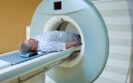
24th October 2022
A gallium positron emission tomography/computed tomography (Ga PET-CT) scan-based radiomics model was better able to distinguish between benign prostate disease and prostate cancer than radiologists according to a retrospective study by Chinese researchers.
Prostate cancer is the 2nd most commonly occurring cancer in men and the 4th most common cancer overall with more than 1.4 million new cases reported in 2020 cancer and 375,304 recorded deaths. Prostate specific membrane antigen (PSMA) is a transmembrane protein that is expressed by virtually all prostate cancers and molecules targeting PSMA can be labelled with radionuclides to become both diagnostic and/or therapeutic agents. Gallium-68 prostate-specific membrane antigen (PSMA) positron emission tomography (PET) has increasingly been utilised globally to assess the local and metastatic burden of prostate cancer. Moreover, a meta-analysis of 37 studies concluded that Ga-68-PSMA PET improves detection of metastases with biochemical recurrence, particularly at low pre-PET PSA levels. A Ga PET-CT scan is normally interpreted by nuclear medicine specialists although there is some evidence to suggest that lesions can be missed. The use of radiomics focuses on improvements in image analysis, using an automated high-throughput extraction of large amounts (200+) of quantitative features of medical images. To date however, the predictive value of a Ga PET-CT scan radiomics model has not been explored for patients with prostate cancer and was the subject of the present study.
The Chinese team retrospectively examined patients who underwent a Ga PET-CT scan and who had pathologically proven prostate cancer or biopsy benign prostate disease. Radiomics prostate cancer features were extracted from the scans and a model created and the ability of the model to differentiate between prostate cancer and benign disease tested. The sensitivity, specificity, positive and negative predictive values of the model were calculated and compared with those of two radiologists who visually inspected the scans.
Ga PET-CT scan radiomics model performance
A total of 125 patients were included and split into a training set (87) and a test set (38). All patients underwent biopsy or surgery and their pathological examination results were assessed.
On the test set, the radiomics model had a sensitivity of 84%, specificity of 77% and and a positive predictive value of 88% for distinguishing between cancer and benign disease.
The agreement between the two radiologists for visual inspection of the scans were very close (kappa = 0.81). However, the corresponding sensitivity for the radiologists on the test set were 74% and specificity was 55% and both were significantly different to the radiomics model (p = 0.036 and 0.002 respectively).
The authors concluded that they had successfully developed and validated a radiomics model based on data extracted from a Ga PET-CT scan and that this non-invasive model was able to predict intraprostatic lesions in men with prostate cancer.
Citation
Zang S et al. Development and validation of 68Ga-PSMA-11 PET/CT-based radiomics model to detect primary prostate cancer EJNMMI Res 2022
IMPORTANT LINKS
MORE FROM COGORA
© Cogora 2025
Cogora Limited. 1 Giltspur Street, London EC1A 9DD
Registered in the United Kingdom. Reg. No. 2147432