This website is intended for healthcare professionals only.
Take a look at a selection of our recent media coverage:
15th January 2025
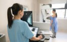
A team from Leeds Teaching Hospitals NHS Trust is leading the national rollout of a new radiographer-led nasogastric tube position check pathway after their efforts showed long-term positive results. Here, the team discusses the need for such a pathway, how it has been shown to improve safety and how other Trusts can get involved.
Over 967,000 nasogastric (NG) tubes are purchased by the NHS each year.1 Less than one in 20 NG tubes are misplaced in the lung, however, failure to recognise this can lead to a never event.2 A ‘never event’ is defined as a ‘serious incident that… (is) wholly preventable’ and results in serious patient harm or death.3
Between 1 April 2014 and 31 March 2024, there were 270 misplaced NG tube-related never events recorded by the NHS in England.4 This has been recognised as a patient safety issue with six national patient safety alerts issued to healthcare providers between 2005 and 2016.1 Despite local systems being implemented and consistent effort from healthcare providers, incidents relating to misplaced NG tubes continue to occur.1
The Health Services Safety Investigation Body (HSSIB), formerly the Healthcare Safety Investigation Branch, is an organisation that investigates patient safety concerns across England to improve patient care. The HSSIB published a report in 2020 on incidents relating to misplaced NG tubes and identified radiographic misinterpretation of NG tube check radiographs as the leading cause of NG tube related never events.1 This is due to the lack of a standardised framework for assessing radiographic interpretation of NG tube radiographs.1
In 2024, a group of professional bodies including the Royal College of Radiologists, British Society of Gastrointestinal and Abdominal Radiologists, the Society and College of Radiographers, and Report and Image Quality Control (RAIQC), collaborated to create a new radiographer-led pathway for utilisation in NHS Trusts and Health Boards across the UK in order to reduce the number of NG tube related never events.
A standardised competency-based training programme was created by the project team and hosted on the web-based platform RAIQC. Here, radiographers are trained to provide contemporaneous radiographic interpretation and to act on their findings. This includes removal of misplaced tubes and arranging for ward staff to attend the department to advance tubes that are not sited far enough into the stomach to be considered safe. This eliminates delays and risk associated with waiting for misplaced tubes to be removed, and, once the tube is correctly sited, allows feeding to be initiated promptly.
This collaborative position check pathway is based on an existing pathway from Leeds Teaching Hospitals NHS Trust, which has been in place for the past 11 years. Empowering the radiographer workforce by expanding their skillset and taking on greater responsibility has prevented further radiograph-related NG tube never events at this multisite Trust since the pathway’s inception.
Regular local audits over these 11 years have demonstrated an accuracy of 99% in the NG tube radiographic interpretation when compared to radiologist review, illustrating the importance of the radiographer workforce in reducing the number of patient safety incidents.
Radiographer training and empowerment to immediately evaluate and act on NG tube check radiographs has produced sustained prevention of patient harm.
The Leeds project team – with support from two radiologists from University College Hospital and Oxford University Hospitals who are involved with the RAIQC platform – is now leading the national roll out of this new radiographer-led pathway project, sharing their experience of utilising the pathway to support implementation at other hospitals.
New and potential site adopters are provided with governance documents and guidance for implementation and encouraged to attend weekly drop-in sessions hosted by the project team for mentoring and support.
Radiologists are key to getting this pathway implemented at Trust level. Any teams interested in adopting it in their service should liaise initially with their clinical lead and contact: [email protected].
Dr Sairah Razak
Radiology registrar
Gillian Roe
Extended imaging practitioner
Dr Damian Tolan
Project lead and consultant gastrointestinal radiologist
Leeds Teaching Hospitals NHS Trust
13th June 2023
A chronic shortage of NHS radiographic professionals, coupled with anticipated surges in demand for heart and kidney scans due to the hot weather, will put patients’ lives at risk this week, the Society of Radiographers (SoR) has warned as it urges members in England to vote for strike action.
Already overstretched radiography teams will be putting in excessive hours to meet a rise in A&E visits caused by the current spell of hot weather, and patients arriving into A&E during this time will have to wait significantly longer for vital scans and diagnosis before their treatment can progress, says the SoR.
Hot weather typically means more cases of heatstroke, heart failure and kidney problems, as well as cuts, sprains, fractures and respiratory problems, which similarly need the attention of radiographic professionals, the Society adds.
Dean Rogers, executive director of industrial strategy and members relations at SoR, said: ‘Doctors and nurses cannot do their jobs without radiographic professionals.
‘Our members are dangerously overstretched. Even when the NHS is not facing increased demands because of the hot weather, nine out of 10 patients will need to see a radiographer, and waiting lists are growing.’
On 19 July 2022 – the hottest UK day on record – there were 638 excess deaths, and 496 excess deaths the following day, according to the Office for National Statistics. The July 2018 heatwave led to a record 2.2 million patients visiting A&E departments in a single month – the highest number since records began in 2010.
Vacancy rates for diagnostic radiographers have risen from 12% to 13% in the last year, and the SoR states that pressure on the radiographic workforce is growing due to widespread training and retention issues.
The SoR is urging its members to vote yes in favour of strike action. Launched on 7 June 2023, its ballot will close at 5pm on 28 June. This follows an indicative ballot in April 2023, in which members voted to reject the Government’s 5% pay offer and non-consolidated lump sum for 2022/23.
Mr Rogers added: ‘We know low pay and poor conditions are forcing radiographers out of the workforce, and they are not being replaced in adequate numbers. Vacancies are running at a minimum of 10 per cent and that’s even with radiographers working considerably more than their contracted hours to ensure that their patients receive the best-possible care.
‘That’s why we’re currently balloting our 20,000 members in England for better pay and working conditions. Every day, the crisis deepens, and this week’s weather will only increase the intolerable pressure on an already overstretched NHS. Our members deserve better. Our patients deserve better.’
Risks to patient safety due to staff shortages have also recently been highlighted by oncology professional organisations in an open letter to the health secretary.
23rd September 2022
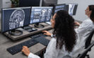
Radiographer diagnostic performance for screening mammography is no different to radiologists for double reading digital mammograms and therefore offers a potential solution to the shortages of radiologists according to the results of a retrospective study by UK researchers.
Screening mammography is widely used in the detection of breast cancer and has been proven to decrease mortality. Moreover, the rate of cancer detection can be further increased by double reading of scans. For example, one study revealed how the relative increase in cancer detection as a result of a second reviewer was 6.3%.
In a 2016 survey, it was found that UK radiographers are already involved with interpreting and reporting images across the full spectrum of clinical indications for mammography including: low-risk population screening, symptomatic, annual surveillance, family history and biopsy/surgical cases. However, despite this change in role, there is limited evidence on the real-life radiographer diagnostic performance in double reading mammograms.
For the present study, the UK team examined the performance of radiographers and radiologists for all screening mammograms in England between 2015 and 2016. The researchers used three key metrics for comparison between radiographers and radiologists: the cancer detection rate (CDR); recall rate (RR) and positive predictive value (PPV) of recall on the basis of biopsy-proven pathological findings for the first readers. Each of the breast scans were analysed based on the reader profession (i.e., radiologist or radiographer) and years of experience.
Radiographer diagnostic performance on screening mammography
A total of 401 readers were included and double read the mammograms of 1,404,395 women. There were 224 radiologists who first read 763,958 mammograms and 177 radiographers who first read 640,437 mammograms.
The overall mean CDR was 7.7 per 1000 examinations and the mean radiographer diagnostic performance was 7.53/1000 examinations and 7.84 for radiologists (p = 0.08). When the researchers analysed CDR’s based on years of experience, there was no variation for either profession (p = 0.87).
The overall recall rate was 5% and again there was no significant difference between radiographers and radiologists (5.2% vs 5%, radiographers vs radiologists, p = 0.63) though the RR was lower for those with more years of experience.
Finally, the overall PPV was 16.7% and again differences between radiographers and radiologists were not significant (16.1% vs 17.1%, radiographers vs radiologists, p = 0.42). As with the RR, PPV improved with more years of experience.
The authors concluded that there were no clear differences in radiographer diagnostic performance and radiologists as readers of screening digital mammograms. They speculated that the use of trained radiographers in the double-reading workflow may offer a potential solution to the shortage of radiologists but suggested that more studies were needed to determine if such physician extender roles can and should, be used to read screening mammograms independent of the radiologist.
Citation
Chen Y et al. Performance of Radiologists and Radiographers in Double Reading Mammograms: The UK National Health Service Breast Screening Program Radiology 2022
1st August 2022
A survey has found that nearly a third of radiographers report being unsure as to how an artificial intelligence system makes its decisions.
In a survey of radiographers, nearly a third stated that they did not know how an artificial intelligence (AI) system made its decisions according to the results of a study by Irish and UK researchers.
A radiology workforce report in 2020 said that across the UK, one in 10 radiologist positions was unfilled. Nevertheless, the reporting of radiographic images by radiographers rather than radiologists is an established practice in the UK and immediate reporting of emergency department radiographs by a radiographer has been deemed a cost-effective service development.
Despite representing a cost-effective service development, as with radiologists, there is a national shortage of radiographers in the UK, with a 2021 report indicating that the average current UK vacancy rate was 10.5% as of November 2020.
To aid both radiologists and radiographers, in recent years there has been an increased use of artificial intelligence (AI) technologies for applications in radiology. Moreover, AI system interpretation of imaging is impressive, with one international study finding that an AI system’s ability to identify breast cancer maintained non-inferior performance and reduced the workload of the second reader by 88%. The introduction of AI systems therefore has the potential to help reduce any backlogs in unreported images.
But the extent to which radiographers understand and interact with AI technology was the subject of the present survey by the Irish and UK team.
They developed a questionnaire focused on AI as used in radiographer reporting and which was initially piloted with a group of 12 radiographers who had a range of different professional backgrounds.
A total of eight questions focussed specifically on AI and its use in radiographer reporting and respondents answered using a seven-point Likert scale (ranging from strongly agree through to strongly disagree). The survey was disseminated via a link posted on professional social media platforms (e.g. LinkedIn and Twitter).
A total of 411 completed surveys responses were received from radiographers working diagnostic and therapeutic radiographer. However, the results of the present study were limited to diagnostic radiographers (86). Perhaps the first and most illuminating finding, was how 89.5% of respondents indicated that they were not currently utilising AI as a part of their reporting role.
In response to a question which asked “I understand how an AI system reaches its decisions” only 28.8% (aggregate value) reported that they ‘somewhat disagreed’, ‘disagreed’ or ‘strongly disagreed’ although 61.6% (aggregate value) that they ‘agreed’ or ‘somewhat agreed’.
In addition, the majority of respondents (59.3%) disagreed that they would be confident in explaining the AI system decision to other healthcare professionals and similarly, only 29.1% agreed that they would be confident explaining the decision to patients or their carers.
While 57% reported that they would feel more certain of their diagnosis if an AI system agreed with their interpretation, the majority (69.8%) stated that they would seek a second opinion if the AI system disagreed with them.
Finally, when asked to rate their trust in an AI system for diagnostic image interpretation on a scale of 1 to 10,, the median score was 5. When asked to choose from a list of possible factors that would increase their level of trust in the AI system, the most common responses were ‘overall performance and accuracy of the system (76%), a visual explanation, such as a heat map (67%) and an indication of the confidence of the AI system in its diagnosis (62%).
The authors concluded that while the majority of respondents were not currently routinely using an AI system as part of their reporting, awareness of how clinicians interact with AI systems was needed as this might promote responsible use of such systems in the future.
Citation
Rainey C et al. UK reporting radiographers’ perceptions of AI in radiographic image interpretation – Current perspectives and future developments Radiography 2022.
IMPORTANT LINKS
MORE FROM COGORA
© Cogora 2025
Cogora Limited. 1 Giltspur Street, London EC1A 9DD
Registered in the United Kingdom. Reg. No. 2147432