This website is intended for healthcare professionals only.
Take a look at a selection of our recent media coverage:
1st June 2022
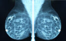
The presence of fatty or dense breasts and benign breast cancer seen on mammography screening in Korean women is associated with a more than two-fold higher risk of future breast cancer. This was the main finding of a retrospective analysis by a team of Korean researchers.
Several factors have been identified as being associated with an increased risk for breast cancer in women which include increasing age, genetics, a family history of the cancer and prior treatment with radiation therapy.
Another factor is the presence of dense breasts upon mammography and one meta-analysis has suggested that the percentage dense area seen on mammography is a strong breast cancer risk factor.
A future factor associated with a higher breast cancer risk is the presence on screening, of both proliferative or non-proliferative benign breast disease, regardless of a family history of breast cancer.
To date, there are limited data on the combined impact of both dense breasts (based on the Breast Imaging Reporting and Data-System (BI-RADS) and the presence of benign breast cancer. One study has indicated that women with benign breast disease and dense breasts are at high risk for future breast cancer.
However, the study was undertaken largely in Westernised populations and there is little information on how the combination of dense breasts and benign breast cancer affect the risk among Asian women, especially given how Asian women have a higher percent breast density compared to Caucasian women.
In the present study, the Korean team retrospectively examined results from a breast screening mammography programme conducted in the Korean population among women 40 years of age and older. These women were followed until the data of their breast cancer diagnosis although individuals with a prior history of breast cancer were excluded.
Breast density was categorised using BI-RADS from A to D based on increasing levels of breast density. The primary outcome of interest was breast cancer and the results were adjusted for several factors such as a family history of the disease, age at menarche, number of children etc.
Dense breasts and breast cancer development
The analysis included 3,911,348 women in whom 58,321 breast cancers developed in women with a mean age of 51.8 years during a median follow-up of 10.6 years.
Breast cancer developed in women with both a higher level of benign breast cancer (18.4% vs 13.3%, p < 0.01) and more dense breasts (55.4% vs 38.4%, p < 0.01) at baseline.
When comparing the different BI-RADs categories, there was an increasing risk of breast cancer with higher breast densities in women who had benign breast cancer.
For example, in women with BI-RADS category A, there was a 49% higher risk of breast cancer in those identified with benign breast cancer (hazard ratio, HR = 1.49, 95% CI 1.40 – 1.58) compared to those in BI-RADS category A but without benign breast cancer.
This risk more than doubled for women with the highest category (BI-RADS D) and benign breast cancer (HR = 2.75, 95% CI 2.63 – 2.88).
The authors concluded that both higher breast density and the presence of benign breast cancer are linked to a higher risk of subsequent breast cancer.
Citation
Kin S et al. Mammographic Breast Density, Benign Breast Disease, and Subsequent Breast Cancer Risk in 3.9 Million Korean Women Radiology 2022
7th April 2022
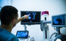
Women having breast tomosynthesis screening over a 10-year period experience a lower cumulative probability of receiving at least one false-positive result compared to conventional mammography. This was according to the results of a study by researchers from the Division of Biostatistics, Department of Public Health Sciences, University of California Davis School of Medicine, California, US.
Screening for breast cancer, even among asymptomatic women, enables the early detection of cancer which is easier to treat than later-stage disease and has a higher probability of survival.
Breast cancer screening is undertaken with mammography and while the technique helps to identify cancer, there is always the risk of a false positive result. In fact, one 10-year study found that one third of women screened had an abnormal test result that required additional evaluation, even though no breast cancer was present.
Other data suggest that that false positive rate is 12% and though the absence of a cancer diagnosis brings relief to women, false-positive findings on screening mammography causes long-term psychosocial harm.
Digital breast tomosynthesis, which approximates a 3D mammogram of the breast, has been found to improve on the cancer detection rate and reduce recall, compared to the more conventional 2-D mammogram. However, effective screening requires women to have many examinations over a period of years and for the present study, the US team wanted to estimate the probability of women receiving at least 1 false-positive result after 10 years of screening with breast tomosynthesis compared to traditional mammography.
They looked at data held within mammography registers covering the period 2005 to 2018 and collected data on self-reported information and the time since the last mammogram from questionnaires. The primary outcomes of interest were false-positive recall, false-positive short-interval follow-up and false-positive biopsy recommendations. The researchers also compared the two approaches based on whether the screening was annual or biennial.
Breast tomosynthesis and false positives
A total of 903,495 women with a mean age at the time of screening of 57.6 years, had 444,704 digital breast tomosynthesis and 2,524,351 mammography examinations. During the period of follow-up, women underwent a mean of 3.3 examinations.
Overall, 7.6% of tomosynthesis and 9% of mammograms resulted in a false-positive recall; 1.8% of tomosynthesis and 2.1% of mammograms led to a false-positive short-interval follow-up recommendation and finally, 1.1% of tomosynthesis and 1.2% of mammograms resulted in a false-positive biopsy recommendation.
The authors calculated that for annual screening the 10-year cumulative probability of at least 1 false-positive with tomosynthesis was 49.6% compared to 56.3% with mammography (difference = -6.7%, 95% CI -7.4 to -6.1) for recall. The cumulative probability of a false-positive for biennial screening was also lower with tomosynthesis compared to mammography (35.7% vs 38.1%).
The researchers concluded that breast tomosynthesis screening led to modest reductions of 2.4% for biennial and 6.7% for annual screening but added how in practise, these percentages equated to thousands of individuals in absolute terms.
Citation
Thao-Quyen HH et al. Cumulative Probability of False-Positive Results After 10 Years of Screening With Digital Breast Tomosynthesis vs Digital Mammography JAMA Netw Open 2022
10th December 2021
The presence of subclinical axillary lymphadenopathy (sLAD) on mammography was significantly more likely among women who had received a COVID-19 vaccination, indicating that they should either delay screening or schedule visits before vaccination. This was the finding of a retrospective study by a team from the Baylor University Medical Center, Dallas, USA.
The term lymphadenopathy refers to lymph nodes that are abnormal in size and axillary lymphadenopathy, describes changes in the size and consistency of lymph nodes in the armpit. There are several potential causes of unilateral axillary lymphadenopathy including breast cancer and which therefore warrants further diagnostic investigations if these changes are observed during screening. Although the presence of sLAD has been observed in women who have received a COVID-19 vaccination, what remains uncertain, is the proportion of vaccinated women that will develop these lesions after either the Pfizer or Moderna vaccines.
Based on case reports of sLAD in vaccinated women, for the present study, the authors investigated the prevalence of the condition in all women who had attended a breast screening programme since December 2020 until April 2021. Data from medical records included the woman’s vaccination status, the vaccine received and the presence or absence of sLAD on mammography. The researchers cross-referenced prior imaging for all women to ensure that there was no baseline evidence of an axillary lymphadenopathy and excluded women who had presented for diagnostic mammography for a palpable axillary lump or pain and those with a history of breast cancer.
Findings
In total, 1027 women with a mean age range of 56.4 to 63.7 years were included in the analysis. There were 158 who received the Moderna vaccine, 144 (Pfizer) and 725 who had not been vaccinated. From these 1027 women, 43 (4.2%) had an sLAD; 3.3% of those who had been vaccinated compared to 0.9% who were unvaccinated and this difference was statistically significant (p < 0.01).
When comparing the two vaccines, the incidence of unilateral axillary lymphadenopathy was higher among those who received the Pfizer vaccination (13.2% vs 9.5%, Pfizer vs Moderna). Among the 43 women with sLAD and who were recalled after screening, vaccine induced sLAD was more likely to have resolved when re-scanned an average of 46.5 days since their COVID-19 vaccination.
The authors concluded that women who have received a COVID-19 vaccination within 8 weeks of their screening mammogram, were significantly more likely to present with a subclinical axillary lymphadenopathy and that this could result in unnecessary further diagnostic workload. They suggested that providers should either delay screening mammograms by 8 weeks in patients who have been vaccinated or that screening should be undertaken before vaccination to avoid unnecessary follow-up.
Citation
Raj S et al. COVID-19 vaccine-associated subclinical axillary lymphadenopathy on screening mammogram Acad Radiol 2021
21st September 2021
Detection of breast cancer requires that mammography scans are interpreted correctly and thus guidelines on screening require that robust quality control and quality assurance are in place. Although an assessment of performance against standards can be achieved through audits, such audits offer little insight of the diagnostic skills of individual radiologists. The use of on-line self-assessment, using pre-diagnosed mammograms, offers a means of identifying any knowledge gaps and could serve as an important educational tool.
An on-line self-assessment tool was adopted by the Italian Society of Medical Radiology in 2018 as part of a national on-line self-assessment program for interpretation of mammograms and the results have published. The test set was a collection of 24 pathology-confirmed cancers combined with 108 mammograms which were reported on as negative after double reading. All cases were acquired with full-field digital mammography and came from women aged 50 – 69 years, who represent those at average risk of breast cancer. For the assessment, the society determined a pass threshold for sensitivity of 62% (amounting to at least 15/24 cancers correctly identified) and 86% specificity (93/108 negative mammograms correctly identified). The on-line self-assessment was posted on a dedicated website and upon registration, radiologists provided information on demographics such as age, gender, place of work (public or private sector), years of breast imaging experience and a qualitative self-judgement of mammography interpretative skills as “beginner”, “average” and “expert”. As with usual mammography practice, the radiologists were required to submit a dichotomous diagnosis (i.e., positive/negative) for every case. In addition to reporting on the radiologist’s success in the test, the authors employed regression analysis and odds ratios to identify the most important predictor variables for diagnostic accuracy.
Findings
A total of 685 radiologists registered for the on-line self-assessment and 49.9% (342) with a mean age of 46 years (69% female), completed test. Among those completing, two-thirds (64%) self-judged their interpretative skills as “average” and respondents had a median of 8 years breast imaging experience although 38.3% reporting having more than 10 years’ experience. Participants reported a median number of 1501 mammographic interpretations per year and the majority (68.7%) worked in the public rather than private sector.
When examining the proportion who successfully completed the assessment, only 28.7% of radiologists (98/342) passed on their first attempt. After an initial failure, 138 of the remaining 244 radiologists, re-took the test of whom, only 35.5% (49/138) passed. In fact, the authors reported that overall, only 44.2% (151/342) of radiologists who completed the on-line self-assessment were successful.
Using regression analysis, a significant association for diagnostic accuracy was found only for the assessment of > 3,000 mammograms per year compared to < 1,000 (Odds ratio, OR = 3.88, 95% CI 1.07 – 14.14, p = 0.04) and working the in public rather than private sector (OR = 1.65). Other variables such as age, self-judged interpretative skills and years of breast imaging experience had no significant effect.
The authors concluded that their study suggested that breast imaging experience does not guarantee diagnostic accuracy in screening reading. They also noted that their on-line self-assessment test could be included as a criterion for the accreditation process of breast units.
Citation
Brancato B et al. Mammography self?evaluation online test for screening readers: an Italian Society of Medical Radiology (SIRM) initiative. Eur Radiol 2021
IMPORTANT LINKS
MORE FROM COGORA
© Cogora 2025
Cogora Limited. 1 Giltspur Street, London EC1A 9DD
Registered in the United Kingdom. Reg. No. 2147432