This website is intended for healthcare professionals only.
Take a look at a selection of our recent media coverage:
29th July 2022
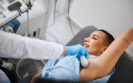
An ultrasound AI system can successfully distinguish between malignant changes in lymph nodes and those due to reactive changes induced after a COVID-19 vaccine, despite the similar appearance, according to the findings of a study by Spanish researchers.
A case of regional lymphadenopathy has been reported following a COVID-19 vaccination as has a case of cervical lymphadenopathy. However, these do not appear to be isolated cases and in a review of 15 studies including 2057 patients, researchers have observed an incidence of lymphadenopathy which ranged between 14.5% and 53% and persisted for longer than 6 weeks in 29% of patients following a COVID-19 vaccination.
The ability to distinguish between the changes present in malignant and benign lymph nodes which have reacted after a COVID-19 vaccination is important, especially in oncology patients. In fact, in a study of five patients, axillary lymphadenopathy occurred after COVID-19 vaccination and the initial radiologic diagnosis raised concerns for metastasis.
In a previous study, the Spanish researchers developed an ultrasound AI system that was able to successfully detect the micro-structural and compositional changes that occur in lymph nodes due to metastatic involvement. As a result, for the present study, the team wondered if they could re-train the ultrasound AI system to differentiate between malignant lymph nodes changes due to metastatic breast cancer (which was used in their previous study) from the reactive changes induced by a COVID-19 vaccine.
The team retrained their ultrasound AI system and tested the model with images containing benign lymph nodes affected by COVID-19 vaccination. They used receiver operating characteristic (ROC) curves to report on the performance of the ultrasound AI system in comparison to expert radiologists (who used a visual scoring method) for the differentiation between the metastatic and benign lymph nodes affected by a COVID-19 vaccination.
Ultrasound AI and radiologist performance
A total of 180 images from 154 patients were used in the analysis. This included 71 images (10 cases and 61 controls) used to re-train the model and 109 (36 cases and 73 controls) as part of the evaluation of the model’s performance.
When comparing the ability of the model and the radiologists to distinguish between metastatic and benign lymph nodes after a COVID-19 vaccination, the ultrasound AI model had an area under the curve of 88.4% compared to 62.6% for the radiologists. This gave a corresponding sensitivity of 77.8% and 41.7% for the ultrasound AI and radiologists respectively.
Furthermore, the ultrasound model had an accuracy of 92.7% compared with 69.7% for the radiologists. In other words, the model had the potential to differentiate between unaffected and metastatic lymph nodes even when both look very similar.
The authors concluded that the ultrasound AI system could be used to differentiate between malignant and benign lymph nodes which changed after COVID-19 vaccination and called for future studies to confirm and validate these preliminary findings.
Citation
Coronado-Gutierrez D et al. Quantitative ultrasound image analysis of axillary lymph nodes to differentiate malignancy from reactive benign changes due to COVID-19 vaccination Eur J Radiol 2022
14th January 2022
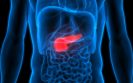
A radiomics nomogram which incorporates the computed tomography (CT) derived radiomics signature and CT-reported lymph node (LN) status, provides favourable preoperative predictive accuracy of LN metastases in patients with pancreatic ductal adenocarcinoma (PDAC). This was the finding from a retrospective study by a team from the Department of Radiology, Changhai Hospital, Shanghai, China.
Pancreatic ductal adenocarcinoma (PDAC) is the fourth-leading cause of cancer related death in the world with a 5-year survival rate of less than 5%. Moreover, pancreatic cancer is a highly lethal disease, for which mortality closely parallels incidence such that every year, more than 350,000 people worldwide are diagnosed and more than 340,000 die of the disease.
The presence of lymph node metastases have been found to be present in up to nearly 68% of patients leading to a significantly poorer prognosis.
The use of multi-slice computed tomography (MSCT) is seen as the best initial diagnostic test for pancreatic cancer although according to a 2014 systematic review, the technique has a low diagnostic accuracy for the detection of LN metastases.
One alternative strategy is the use of radiomics, which represents a quantitative and non-invasive approach to imaging and aims to enhance the existing data to clinicians by means of advanced mathematical analysis. While the use of radiomics has been found to be of value in detection of some cancers, it has not been used for predicting LN metastases in those with PDAC.
For the present study, the Chinese team, aimed to develop and validate a radiomics nomogram that incorporated a radiomics signature and CT-reported LN status for the pre-operative prediction of LN metastasis in those with PDAC.
Patients who had undergone MSCT were divided into a training and validation cohort with both groups including LN negative and positive individuals. The authors used multivariable logistic regression analysis to develop a model to predict LN metastases and the area under curve (AUC) values used to estimate the model’s sensitivity, specificity and accuracy.
Findings
A total of 225 patients aged between 59 and 64 years of age were split into a training (180) and validation cohort (45). Using univariate analysis, only the radiomics score (rad-score) (p < 0.0001) and CT-reported LN status (p = 0.014) were significantly associated with an increased risk of LN metastases.
In the validation cohort, the radiomics model yielded an AUC of 0.81, giving a sensitivity of 84.2%, a specificity of 69.2% and an accuracy of 75.6%. Using just the radiomics score and CT-reported LN status, decision curve analysis showed that with the nomogram, if the threshold probability was between 0.25 and 0.75, using the nomogram to predict LN metastases added more benefit that a treat-all patients strategy.
They concluded that the radiomics nomogram showed favourable accuracy for the pre-operative prediction of LN metastases in PDAC patients.
Citation
Bian Y et al. Radiomics nomogram for the preoperative prediction of lymph node metastasis in pancreatic ductal adenocarcinoma. Cancer Imaging 2022.
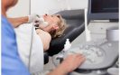
Ultrasonography (US) is better than clinical examination for the detection of lymph node (LN) metastases in patients with squamous cell carcinoma (SCC) but is associated with higher rate of false positives. This was the conclusion of a study by a team from the Department of Dermatology, Erasmus Medical Center Cancer Institute, Rotterdam, the Netherlands.
Squamous cell carcinoma represents a malignant proliferation of the cutaneous epithelium and accounts for 20% to 50% of skin cancers. Fortunately however, the incidence of metastases with the cancer is low, with one UK study observing that after 36 months, 1.1% of cases in women and 2.5% in men become metastatic. Although a 2020 interdisciplinary European consensus guideline noted that imaging methods, for example, US are more sensitive than clinical examination, it added that ‘there are limited data on the use of US for nodal metastasis‘ for SCC.
Where clinical examination reveals suspicious findings, clinicians may request US although the added value of this imaging modality remains uncertain. For the present analysis, the Dutch researchers therefore examined the diagnostic accuracy of clinical examination and baseline US for the detection of LN metastases in patients with high risk cutaneous SCC of the head and neck. They also explored the accuracy of baseline US where baseline clinical examination did not suspect metastases.
The team retrospectively reviewed all consecutive, high risk patients at their centre. Where lymphadenopathy was detected upon examination or with one or more suspicious lymph nodes detected on US, a biopsy was undertaken and nodal metastasis was confirmed by cytology. The diagnostic accuracy of clinical examination and US was calculated based on sensitivity, specificity and positive predictive value (PPV).
Findings
During a 3-year period, 233 patients with a median age of 79.1 years (75.5% male) with 246 high-risk SCCs were included in the analysis.
Among the 246 tumours, 20 (8.1%) had suspicious lymphadenopathy upon clinical examination and 11 confirmed as LN metastatic after cytology. Suspicious lymph nodes were found on US in 28.5% (70/246) of cases and in 20 of these, metastasis was confirmed by cytology.
Using these data, the authors calculated the sensitivity of clinical examination to be 50% (95% CI 28 – 72%), with a specificity of 96% (95% CI 93 – 98%) and a PPV of 55%. In contrast, US had a sensitivity of 91% (95% CI 71 – 99%), a specificity of 78% (95% CI 72 – 83%) and a PPV of 29%.
In patients with a negative result upon clinical examination, 9 of 11 metastases were detected by US with a sensitivity of 82% although there were 45 false positives, making the PPV only 17%. Based on these findings, the authors suggested that while US examination was very sensitive, this needs to be seen in the context of a higher number of false positives.
The concluded that while US was a more sensitive screening tool to detect LN metastasis, the high false positive rate would in practice lead to a higher rate of unnecessary US and cytology procedures. They suggested that future studies should focus on identifying specific populations of those with SCCs who would benefit the most from baseline US to detect metastatic disease.
Citation
Tokez S et al. Assessment of the Diagnostic Accuracy of Baseline Clinical Examination and Ultrasonographic Imaging for the Detection of Lymph Node Metastasis in Patients With High-Risk Cutaneous Squamous Cell Carcinoma of the Head and Neck JAMA Dermatol 2021.
IMPORTANT LINKS
MORE FROM COGORA
© Cogora 2025
Cogora Limited. 1 Giltspur Street, London EC1A 9DD
Registered in the United Kingdom. Reg. No. 2147432