This website is intended for healthcare professionals only.
Take a look at a selection of our recent media coverage:
16th December 2022
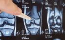
Non-steroidal anti-inflammatory drugs (NSAIDs) used for the management of osteoarthritis appear to worsen inflammation in the knee joint over time, according to a study presented at the Radiological Society of North America (RSNA) conference 2022.
NSAIDs such as ibuprofen and naproxen are widely recommended and prescribed to treat pain in osteoarthritis yet are also known to elicit adverse events affecting the gastrointestinal, cardiovascular, and renal systems. In fact, one US study of patients with knee osteoarthritis (OA) revealed how non-prescription non-steroidal anti-inflammatory drugs were the most frequently reported medications (26.8%), even in those more than 75-years old. In recent years attention has focused on the role of the synovium in OA and how the presence of synovitis is associated with more severe pain and joint dysfunction and may be predictive of faster rates of cartilage loss in certain patient populations. However, whether NSAIDs are beneficial to patients with OA in the longer term remains uncertain, though some data suggests that there may not be long-term benefits given how a majority of patients had persistent pain and disability despite therapy.
In the study presented at RSNA, researchers set out to analyse the association between NSAID use and synovitis in patients with osteoarthritis of the knee and to assess how treatment with NSAIDs affects joint structure over time. The team used data derived from participants from the Osteoarthritis Initiative (OAI) cohort who had moderate to severe OA and sustained NSAID treatment for at least 12 months between baseline and 4-year follow-up. The outcomes for these participants were then compared with non-NSAID treated participants. All participants underwent a 3T MRI of the knee at baseline and after 4 years. and images were semi-quantitatively scored for MR biomarkers of synovial inflammation (effusion-synovitis, size and signal intensity of infra-patellar fat pad (IFP) and the synovial proliferation score (SPS). Cartilage thickness and T2-relaxation time measurements served as non-invasive biomarkers for evaluating OA progression. The associations between baseline and findings after 4 years were investigated with linear regression models (including adjustment for sex, BMI, age, pain, K/L grade).
NSAIDs and arthritic markers over time
A total of 721 participants (129 with and 592 participants without regular usage of NSAID) were included in the analysis. At baseline, there was a significantly higher signal intensity in the IFP among NSAID users as compared to controls (adjusted difference in score = 0.26, 95% CI -0.5 – 0.129, p = 0.039). Additionally, when assessed over time, there was a significantly higher increase in the signal intensity of IFP (0.46, 95% CI 0.2 – 0.72, p < 0.001) and a higher increase in effusion synovitis (0.27, 95% CI 0.06 – 0.47, p = 0.01) in NSAID users compared to controls. The size of IFP and SPS did not show a significant difference between groups at baseline and no significant change over time. NSAID users showed more degenerative changes regarding T2-relaxation time and cartilage thickness over time, but this did not reach statistical significance.
Based on these findings, the authors suggested that among those using NSAIDs, there was a higher signal intensity in IFP and more effusion/ synovitis than controls, indicating that long-term NSAID usage is associated with more synovitis.
Commenting on these findings in a press release, lead author, Johanna Luitjens, a postdoctoral scholar in the Department of Radiology and Biomedical Imaging at the University of California, San Francisco, said ‘in this large group of participants, we were able to show that there were no protective mechanisms from NSAIDs in reducing inflammation or slowing down progression of osteoarthritis of the knee joint.’ She added that ‘the anti-inflammatory effect that normally comes from NSAIDs may not effectively prevent synovitis, with progressive degenerative change resulting in worsening of synovitis over time.’
Citation
Luitjens J et al. Impact of Non-steroidal Anti-inflammatory Drugs (NSAIDs) on Synovitis and the Progression of Osteoarthritis: Data from the Osteoarthritis Initiative (OAI). RSNA conference 2022.
21st November 2022
Using an MRI derived radiomics nomogram that also includes clinical patient characteristics helps to predict which patients with knee osteoarthritis are likely to see improvements in their pain over a 2-year period according to a study by Chinese researchers.
Osteoarthritis (OA) is a common condition that affects 7% of the global population which amounts to more than 500 million people. Although OA has been assessed conventionally using X-rays, an alternative is magnetic resonance imaging (MRI). In fact, it has been suggested that MRI is the best modality for imaging of osteoarthritis enabling visualisation of multiple individual tissue pathologies relating to pain and also to predict clinical outcome. When Chinese researchers undertook a randomised trial to examine the effect of vitamin D on OA knee pain, compared with placebo, there were no significant differences in MRI-measured tibial cartilage volume or knee pain score over 2 years. However, in a post-hoc analysis, 64% of vitamin D participants 57% of placebo participants achieved a 20% improvement in knee pain score over 2 years.
Give that a fifth of participants had actually seen an improvement in their knee pain, researchers wondered how it might be possible to identify those patients who were likely to benefit from vitamin D. They decided to create a radiomics nomogram based on MRI-derived features of subchondral bone together with clinical factors and which could be used to predict an improvement over 2 years in knee OA pain. The team used data from the VIDEO study of knee osteoarthritis in which participants had an MRI scan at baseline and after 24 months. The primary outcome was knee pain, and which was assessed using the WOMAC score. The team used MRI data to create a radiomics model which was trained and then validated with separate cohorts from the VIDEO trial. The data from the model was then used to produce a nomogram for predicting osteoarthritis knee pain improvement over two years. The model was assessed based on the area under the receiver characteristics operating curve (AUC).
MRI derived radiomics model and prediction of knee pain
A total of 216 patients with mean age of 68.3 years (47% female) were included and 172 were used in the training cohort, of whom, 78 had no improvement in pain and the remainder used in the validation cohort.
Only two variables, female gender and baseline total knee pain score were significant predictors of an improvement in knee pain over 2 years and were used in the clinical model, together with vitamin D supplementation. The MRI derived model included a radiomics signature and the two significant clinical variables.
In the validation cohort, the nomogram had a higher AUC than the clinical model (0.83 vs 0.71) for the prediction of knee pain improvement although this difference was not significant (p = 0.08). In addition, the use of a decision curve analysis confirmed the clinical usefulness of nomogram.
The authors concluded that their radiomics-based nomogram which comprised the MR radiomics signature and clinical variables achieved a favourable predictive efficacy and accuracy in differentiating improvement in knee pain among OA patients.
Citation
Lin T et al. Prediction of knee pain improvement over two years for knee osteoarthritis using a dynamic nomogram based on MRI-derived radiomics: a proof-of-concept study. Osteoarthritis Cartilage 2022
21st July 2022
The use of intra-articular hyaluronic acid (HA) produces a small, significant increase in knee osteoarthritis pain intensity compared to placebo although this reduction is less than the minimal clinically important difference according of a systematic review and meta-analysis by Swiss and Canadian researchers.
Osteoarthritis is a degenerative condition and which globally affects around 16% of those aged 15 and over but 22⋅9% of individuals aged 40 and over. One form of treatment is viscosupplementation with intra-articular hyaluronic acid and although it provides a clinically meaningful benefit to a large number of patients, emerging evidence indicates that this is largely a result of other factors, including the placebo effect.
With its widespread use yet apparent lack of evidence, for the present study, the research team decided to perform a systematic review and meta-analysis to examine the evidence on the clinical benefits and safety of the intervention.
The team searched for randomised or quasi-randomised trials in which at least 75% of participants had clinically or radiologically confirmed knee osteoarthritis and where outcomes such as pain, function or adverse events were reported. The primary outcome was set as pain intensity with function and serious adverse events as the two secondary outcomes.
Continuous outcomes were analysed as standardised mean differences (SMDs) such that when the SMD was > 0, this was indicative of a better outcome for HA. The researchers also determined that the minimal clinically important difference for HA was -0.37.
Hyaluronic acid and pain intensity
A total of 169 trials with 9,423 participants and a mean age of 62 years (59% women) and with a mean disease duration of 5.2 years were included in the analysis. The median follow-up time for patients after their HA injection was 13 weeks and 12 weeks for functional assessment.
Based on 24 trials (8997 randomised patients), there was a small, but significant though non-clinically relevant reduction in pain intensity after HA injection (SMD = -0.08, 95% CI -0.15 to -0.02, p = 0.02). Based on a 100 mm visual analogue pain scale, this equated to a 2 mm reduction in scores compared to placebo.
For the secondary outcome of function, the pooled estimate was a SMD of -0.11 (95% CI -0.18 to -0.05, p = 0.001) which again was much lower than the minimally important difference.
In terms of adverse effects, the use of HA was associated with a significant increased risk of serious adverse events compared to placebo (relative risk = 1.49, 95% CI 1.12 – 1.98, p = 0.003). Overall, 3.7% of patients receiving HA and 2.5% of those given a placebo experienced a serious adverse event.
The authors concluded that HA was associated with a clinically irrelevant reduction in pain intensity and suggested that their findings do not support the broader use of this intervention for knee osteoarthritis.
Citation
Pereira TV et al. Viscosupplementation for knee osteoarthritis: systematic review and meta-analysis BMJ 2022
IMPORTANT LINKS
MORE FROM COGORA
© Cogora 2025
Cogora Limited. 1 Giltspur Street, London EC1A 9DD
Registered in the United Kingdom. Reg. No. 2147432