This website is intended for healthcare professionals only.
Take a look at a selection of our recent media coverage:
9th September 2022
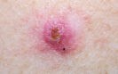
Basal cell carcinoma can be diagnosed more accurately than with either clinical or dermoscopic examination using a novel imaging technique according to data presented at the 31st European Academy of Dermatology and Venereology (EADV) Congress by researchers from the Department of Dermatology at the Hôpital Erasme, Université Libre de Bruxelles, Brussels.
Basal cell carcinoma (BCC) is the most frequently diagnosed malignancy worldwide and a Dutch study found that over the period 2001-2019, the age-standardised incidence rates for both men and women with a first BCC increased from 157 to 304 and from 124 to 274 per 100 000 person-years, respectively.
Furthermore, the authors of the study concluded that ‘the incidence continues to rise in patients aged 50 years and older. In the next decade a further increase in BCC incidence is expected.’
According to the European Skin Cancer Foundation, in Europe the incidence is of BCC is about 50 to 80 new patients per 100.00 persons per year but which is less than in Australia, where the incidence is about 250 per 100.00 persons per year and with upward trend.
In the current study, researchers found that using a new, non-invasive skin imaging technology called line-field confocal optical coherence tomography (LC-OCT), which gives detailed 3D images at cellular level, significantly increased diagnostic accuracy.
For the differentiation of BCC from BCC-imitators (such as squamous cell carcinoma, actinic and seborrheic keratosis, dermal nevus, and inflammatory conditions), the researchers analysed 303 lesions, including 173 BCC and 130 BCC-imitators.
They found that LC-OCT significantly increased the diagnostic accuracy by 12% compared to dermoscopic examination alone (from 85% up to 97%), which the most commonly used skin cancer diagnostic technique.
Importantly, for the differentiation of superficial BCC (a subtype that can be treated non-surgically) from other BCC subtypes, using LC-OCT again increased the diagnostic accuracy by 12% compared to dermoscopic examination alone (from 80% to 92%).
The study also produced a diagnostic algorithm useful to guide the clinician’s diagnosis towards different BCC and BCC-imitators’ subtypes. The algorithm is based on the most powerful LC-OCT morphological criteria that came out from their comprehensive statistical analysis.
Professor Mariano Suppa, a lead researcher and consultant dermatologist from Italy said: ‘Our findings suggest that, when in front of an BCC equivocal lesion, LC-OCT enables a more accurate diagnosis and, therefore, should be included in the diagnostic process and management of BCC.’
Prof. Suppa added: ‘LC-OCT has the potential to reduce the number of unnecessary biopsies and excisions in cases of superficial BCC and also in the case of benign lesions that do not require surgery.’
14th January 2022
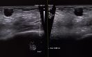
Diagnostic ultrasound appears to be the imaging of choice for an assessment of intra-operative margin assessment after excision of breast cancer according to a meta-analysis by researchers from Aarhus University Hospital, Denmark.
According to the World Health Organisation, in 2020 there were 2.3 million women diagnosed with breast cancer and globally, 685 000 deaths. The primary treatment modality for breast cancer is surgery with the aim of removing the cancer and conserving as much healthy tissue as possible.
However, there is currently a lack of consensus of an adequate margin in patients with invasive or in situ breast carcinoma undergoing breast-conserving surgery and over 20% of women require reexcision following their initial surgery.
Given the current reexcision rates, there is an obvious and important need for techniques to assess intra-operative breast margins. While frozen section and cytology have been shown to have the greatest diagnostic accuracy when assessing intra-operative breast margin assessment, such methods are resource intensive and turnaround times for results have prevented widespread adoption.
Although optical methods to determine margin status have been developed and with a high sensitivity and selectivity, no current modality has matured into a complete solution to the margin problem.
For the present study, the Danish team sought to evaluate the pooled diagnostic sensitivity, and specificity of radiological methods for intra-operative margin assessment and their impact on repeat surgery rate.
They performed a systematic search in PubMed, Embase, Cochrane Library, Scopus, and Web of Science for studies using several imaging techniques including radiography, digital breast tomosynthesis (DBT), micro-CT, and diagnostic ultrasound and where the histological assessment was used as the reference method.
The area under the curve (AUC) for each technique was calculated and a bivariate random effect model was used to obtained pooled sensitivity and specificity of the index tests in the meta-analysis.
Findings
The systematic search resulted in 22 articles with 29 radiological imaging methods and were included in the meta-analysis. Pooled sensitivity and specificity and area under the curve were calculated for a total of four subgroups: radiography, DBT, micro-CT and diagnostic ultrasound.
The AUC for radiography was 60%, DBT 76%, micro-CT 72% and diagnostic ultrasound 80%. The sensitivities were 52%, 67%, 68% and 72% for radiography, DBT, micro-CT and diagnostic ultrasound respectively. The corresponding specificities were 77%, 76%, 72% and 80%. However, the repeat surgery rate was poorly reported in the studies.
Although based on these findings, diagnostic ultrasound appeared to be the best imaging modality, the authors felt that use of ultrasound was not realistic for the current workflow processes and concluded that the ‘radiological methods for margin assessment need further improvement to provide reliable guidance in the clinical workflow and to prevent repeat surgeries.’
Citation
Manhoobi IP et al. Diagnostic accuracy of radiography, digital breast tomosynthesis, micro-CT and ultrasound for margin assessment during breast surgery: A systematic review and meta-analysis. Acad Radiol 2022.
15th October 2021
Understanding of medical radiation appears to be limited among patients undergoing various imaging modalities according to a survey by a team from the department of Diagnostic and Interventional Radiology, University of Pisa, Italy. The use of imaging techniques have become increasingly important over the years as a diagnostic aid to clinicians although concerns have been raised, especially among the paediatric population, that greater use of modalities such as computed tomography (CT), increase exposure to radiation. Moreover, there is some evidence that among individuals aged under 19 years of age, the overall cancer incidence was 24% higher in those exposed to radiation from CT scans.
But how much do patients who undergo different imaging modalities understand about medical radiation and its associated risks? This was the question addressed by the Italian team who undertook a survey among patients awaiting a medical imaging procedure at 16 hospitals between June 2019 and May 2020. The survey included 23 questions which comprised three sections: the first covered patient demographics including levels of education, area of residence; the second, knowledge about ionising radiation risks and the final section enquired about communication of the risks and current sources of information on these risks.
Findings
The medical radiation survey was completed by 3039 individuals with a mean age of 44.9 years (53.4% female) with virtually all respondents (98.5%) having previously undergone at least one radiological test in their life. In terms of knowledge about the different imaging modalities, ultrasonography was correctly identified as a radiation-free technique by 85% of respondents. Mammography was correctly identified as an ionising modality by only 38.4% but 71% were correct in recognising that CT scans were also ionising. In contrast, however, only 43% of respondents knew that MRI was non-ionising and more than half (55.1%) were unaware that chest CT scans delivered a larger dose of radiation than chest radiography.
Perhaps of more concern, was how nearly half (44.4%) deemed their knowledge of radiation risks as inadequate with only a quarter (24.9%) reporting it as being either fair/good or excellent. Information sources about the risks associated with ionising radiation included radio/television (27.6%), the internet/social media (25.3%) but 35% stated that they had never received any such information. Interestingly, only 42.6% of respondents had been informed about radiation risk during a previous imaging scan and the majority of respondents (80.4%) expressed a preference to receive such information from their healthcare professionals.
The authors concluded that their survey suggested a substantial lack of knowledge about medical radiation and called for improved communication between medical staff and patients to increase awareness of the topics and risks from cumulative exposure.
Citation
Bastiani L et al. Patient Perceptions and Knowledge of Ionizing Radiation From Medical Imaging. JAMA Netw Open 2021
IMPORTANT LINKS
MORE FROM COGORA
© Cogora 2025
Cogora Limited. 1 Giltspur Street, London EC1A 9DD
Registered in the United Kingdom. Reg. No. 2147432