This website is intended for healthcare professionals only.
Take a look at a selection of our recent media coverage:
6th May 2022
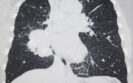
The detection of emphysema via visual or quantitative assessment on a CT-scan has been found to be linked with a higher odds of developing lung cancer.
This was the conclusion of a systematic review by researchers from the Departments of Epidemiology, Radiology and Pulmonary Diseases, University Medical Center Groningen, University of Groningen, Groningen, the Netherlands.
A chest computer tomography (CT) scan enables quantification of the amount of emphysema present in the lungs and while some evidence suggests that emphysema on a CT scan is related to lung cancer in a high-risk population, other data indicates that no CT measures of emphysema have an independent association with lung cancer.
With some uncertainty over the association between the presence of emphysema seen on a CT scan and lung cancer, for the present study, the researchers decided to undertake a systematic review and meta-analysis to further probe this association.
They searched all the major databases and included studies that specifically assessed the association between emphysema and the diagnosis of lung cancer based on histopathologic examination.
The team defined visual emphysema as disrupted lung vasculature and parenchyma with low attenuation occupying any lung zone on the chest CT scan and quantitative emphysema as the percentage of total lung volume below a given Hounsfield unit threshold (-950 HU at full inspiration). They also sought to examine whether the severity of emphysema was associated with lung cancer and graded this as trace, mild, moderate/severe.
The studies were stratified based on whether visual or quantitative assessments were used and the presence of confirmed lung cancer was the main outcome of interest expressed and expressed as an odds ratio, adjusted for age, gender and smoking status.
Emphysema and lung cancer risk
A total of 21 studies met the inclusion criteria with 3907 patients who had lung cancer and 103,175 controls.
The pooled odds ratio (OR) for lung cancer in the presence of emphysema was 2.3 (95% CI 2 – 2.6) in studies which employed visual assessment and 2.2 (95% CI 1.8 – 2.8) where the authors used quantitative assessment.
When stratified by disease severity, the overall pooled OR for lung cancer increased with disease severity although there were differences based on whether the data was acquired by visual or quantitative assessment.
For example, in studies that employed visual assessment, the ORs for lung cancer were 2.5 (trace disease), 3.7 (mild disease) and 4.5 (moderate to severe disease).
While these odds ratios were still elevated based on quantitative assessments, the magnitudes were slightly lower e.g., 1.9 for trace disease and 2.5 (moderate to severe disease).
Based on their findings, the author concluded that the presence of emphysema diagnosed on a chest CT scan was independently associated with a higher odds of developing lung cancer.
Citation
Yang X et al. Association between Chest CT–defined Emphysema and Lung Cancer: A Systematic Review and Meta-Analysis Radiology 2022
15th November 2021
Using dark field x-ray, a team from the Department of Physics, Munich School of BioEngineering, Germany, were able to diagnosis emphysema in patients with COPD which to date has not been possible with conventional x-ray methods. Emphysema is caused by the irreversible destruction of alveolar walls, leading to enlargement of distal airspaces. Although this leads to changes in the lung structure, it is not possible to detect the early stages of emphysema with a conventional chest x-ray. As x-rays are subject to other effects such as refraction and ultra small-angle scattering which are not visualised with a conventional X-ray imaging system, in DFX, the contrast is produced by these multiple refractions on microstructures in the object. Hence, the dark-field signal enables visualisation of structural information that is inaccessible with conventional medical x-ray systems and dark field imaging has, for example, enabled the identification of acute lung inflammation in animal models.
With the potential to identify changes at the micro-structural level with dark field x-ray, indicative of emphysema, the German team undertook their study to examine whether this approach could improve the medical lung assessment in patients with COPD. Included patients were those with an initial indication of emphysema as revealed by a CT scan but still absent based on spirometry readings. However, a small number of patients with moderate to severe emphysema were also included. COPD classification was based on the post forced expiratory volume in one second (FEV1) and the forced vital capacity (FVC) as proposed by GOLD classification and patients with a FEV1/FVC value of 0.70 were assessed as having no COPD. Dark field x-rays were performed and visually assessed by five readers.
Findings
A total of 77 patients with a mean age of 65.9 years (male) and 63.3 years (female) were recruited with the majority (83%) at GOLD stage 0. Focusing on the first patient examined, the researchers described how the DFX showed that most parts of the lung yielded dark field values comparable with a healthy lung. A group of 42 patients from the cohort underwent diffusion capacity testing and it was found that the dark field signal gave a strong correlation than with either the CT emphysema index or the visual emphysema grading based on the CT-images.
Commenting on their findings, the authors reported that “in direct comparison, dark-field images and visual evaluation of CT images yield consistent findings regarding emphysema diagnosis.” They also added that the visual features seen with the dark field appeared to provide a greater diagnostic value than conventional emphysema charactering parameters. Moreover, while noting that there are currently no commercially available dark field systems available, their study has shown how the system does not require specialist knowledge and that it is operationally comparable to conventional radiography systems.
In their conclusion, they noted how DRX could offer a low radiation alternative to CT in patients with COPD adding that “x-ray dark-field chest imaging could contribute to improving the detection, diagnosis, and thus treatment and care of pulmonary disorders.”
Citation
Willer K et al. X-ray dark-field chest imaging for detection and quantification of emphysema in patients with chronic obstructive pulmonary disease: a diagnostic accuracy study. Lancet Digit Health 2021
IMPORTANT LINKS
MORE FROM COGORA
© Cogora 2025
Cogora Limited. 1 Giltspur Street, London EC1A 9DD
Registered in the United Kingdom. Reg. No. 2147432