This website is intended for healthcare professionals only.
Take a look at a selection of our recent media coverage:
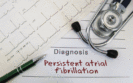
18th November 2022
A model combining features of an ECG and radiomics data derived from cardiovascular magnetic resonance imaging improved the detection of atrial fibrillation (AF) in women to a greater extent than either model alone according to the findings of a study by Spanish and UK researchers.
Atrial fibrillation is the most common cardiac arrhythmia, characterised by an irregular heart rhythm and an often abnormally rapid heart rate and globally has been estimated to affect 0.51% of the population. AF is diagnosed from an ECG in which typically, P-waves are absent and there is both a chaotic baseline and an irregular ventricular rate. Cardiac magnetic resonance (CMR) is the reference imaging modality for assessment of cardiac structure and function and CMR radiomics has the potential to improve diagnostic accuracy. Moreover, Cardiac MRI of the atrial substrate is not only a tool for management and treatment of arrhythmia, but also to individualise the prevention of stroke and major cardiovascular events. But what remains unclear is whether the use of a CMR-based radiomics model can identify patients with AF and more importantly, if addition of a model that uses ECG-derived data, would further enhance the potential to detect AF.
In the present study, researchers examined the feasibility of combining a CMR-derived radiomics model and one based on ECG data. The team used information from the UK Biobank and identified patients who had both an ECG and CMR scan and compared their findings with healthy controls. Models were assessed using the area under the receiver operating characteristics curve (AUC) and associated sensitivity and specificity.
ECG and radiomics model and atrial fibrillation detection
A total of 32,121 participants with a mean age of 63 years (51% female) were included and of whom, 495 (63% male) had AF.
Overall, the AUC for the combined model was similar to the ECG-model (0.87 vs 0.86), i.e., adding the radiomics model did not significantly improve predictive power. In fact, when comparing the predictive power of models between the sexes, the AUC for the ECG-model was less predictive for women than men (0.77 vs 0.88, p < 0.05). However, although accuracy improved for women when combined with the CMR-model, but this only improved to the level of the ECG-model for men (0.87 vs 0.88, women vs men). Finally, when considering AF patients who had a normal ECG, the combined model had an AUC of 0.61.
The authors concluded that their integrative radiomics-ECG model presents a potential novel approach for earlier detection of AF.
Citation
Pujadas ER et al. Atrial fibrillation prediction by combining ECG markers and CMR radiomics. Sci Rep 2022
6th December 2021
The presence of abnormal axis and left bundle branch block (LBBB) on an electrocardiogram (ECG) have been found to be independent predictors of in-hospital mortality among patients with COVID-19, according to researchers from the Department of Emergency Medicine, Nantes, France.
It has been already established that almost all patients (93%) who are critically ill with COVID-19 will show changes on their ECG. However, given that these data were obtained from those who were most ill, there is some degree of uncertainty whether such findings are generalisable to those with less severe COVID-19.
For the present study, the US team set out to retrospectively examine whether there were any specific ECG patterns or changes that could be associated with in-hospital mortality from COVID-19. The study was conducted in three French hospitals in adults (>18 years of age) and who presented to an emergency department, with a PCR confirmed infection with COVID-19 and for whom an ECG was undertaken. The team collected demographic details and co-morbidities and set the primary outcome as in-hospital mortality. The ECGs were independently assessed by two emergency care physicians (one of whom was a cardiologist) who documented rhythms, the presence of atrial fibrillation and any abnormal axis changes and conduction blocks.
Findings
A total of 275 patients with a mean age of 70 years (56% male) had an ECG upon admission to the emergency department. Overall, 21% of patients had chronic kidney disease, 13% diabetes and 14% cardiovascular disease. Interestingly, 13% of those presenting at the department reported chest pain and 2%had palpitations. From the 275 patients, 14% died at hospital.
Among those who survived (n = 238) 89% were in sinus rhythm compared with 12% of those who died. An abnormal axis was rare and observed in only 6% of the cohort and LBBB in 4%. However, both changes were higher among those who subsequently died compared to survivors (14% (both abnormalities) vs 4% and 3% respectively).
Using univariate and then multivariate regression analysis, only an abnormal axis and LBBB were significantly associated with in-hospital mortality. For an abnormal axis, the adjusted odds ratio, OR was 3.9 (95% CI 1.1 – 11.5, p = 0.02) and for LBBB the OR was 7.1 (95% CI 1.9 – 25.1, p = 0.002).
Commenting on their findings, the authors emphasised the importance of detecting for cardiac impairment upon admission to an emergency department and in particular, an abnormal axis and LBBB. Nevertheless, they recognised that due to the retrospective nature of their study, it was not always possible to determine if the ECG abnormalities were due to COVID-19 or a prior cardiopathy.
They concluded by calling for a prospective study to evaluate ECG monitoring in COVID-19 prognosis and whether the specific abnormalities that they detected during infection with the virus, constitute a risk factor for a subsequent cardiovascular event.
Citation
De Carvalho H et al. Electrocardiographic abnormalities in COVID-19 patients visiting the emergency department: a multicenter retrospective study BMC Emerg Med 2021
IMPORTANT LINKS
MORE FROM COGORA
© Cogora 2025
Cogora Limited. 1 Giltspur Street, London EC1A 9DD
Registered in the United Kingdom. Reg. No. 2147432