This website is intended for healthcare professionals only.
Take a look at a selection of our recent media coverage:
21st November 2024
A new survey by the European Society of Paediatric Research (ESPR) has examined how widely lung ultrasound is used in neonatal intensive care units (NICUs) across Europe to better understand what it is used for and how this may be improved.
The survey results, published on behalf of the ESPR Pulmonary Research Consortium, found that although lung ultrasound is available in NICUs throughout the continent, uptake is highly variable.
To improve implementation, the authors suggest the development of learning opportunities for healthcare professionals, as well as the establishment of international guidelines.
The researchers analysed lung ultrasound use in NICUs using an international online survey undertaken in 2023, collecting data from 560 NICUs in 24 countries.
The percentage of NICUs using this technique varied widely between countries, ranging from 20% to 98%. Of the NICUs surveyed, 76% of the units used it for patient care, while 6% used it only for research purposes.
Where lung ultrasound was used in a clinical context, it was most frequently used to diagnose respiratory diseases (68%), to evaluate an infant experiencing acute clinical deterioration (53%) and to guide surfactant treatment (39%).
Respiratory conditions diagnosed by in this way most commonly included pleural effusion, pneumothorax, newborn transient tachypnea and respiratory distress syndrome.
In all NICUs, lung ultrasound was mainly used by neonatologists. The researchers found that experience using it varied widely across Europe, with only 13% of the units having more than five years of experience using the machines. One-third of the units had less than two years’ experience.
The most common reasons for not using lung ultrasound were a lack of technical experience and uncertainty around image interpretation.
Survey respondents and authors suggested that specific courses and an international guideline on neonatal lung ultrasound could promote the uptake of this technique.
Reference
Alonso-Ojembarrena, A et al. Use of neonatal lung ultrasound in European neonatal units: a survey by the European Society of Paediatric Research. Archives of Disease in Childhood. Fetal and Neonatal Edition 2024; Apr 11: DOI: 10.1136/archdischild-2024-327068.
14th January 2022
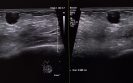
Diagnostic ultrasound appears to be the imaging of choice for an assessment of intra-operative margin assessment after excision of breast cancer according to a meta-analysis by researchers from Aarhus University Hospital, Denmark.
According to the World Health Organisation, in 2020 there were 2.3 million women diagnosed with breast cancer and globally, 685 000 deaths. The primary treatment modality for breast cancer is surgery with the aim of removing the cancer and conserving as much healthy tissue as possible.
However, there is currently a lack of consensus of an adequate margin in patients with invasive or in situ breast carcinoma undergoing breast-conserving surgery and over 20% of women require reexcision following their initial surgery.
Given the current reexcision rates, there is an obvious and important need for techniques to assess intra-operative breast margins. While frozen section and cytology have been shown to have the greatest diagnostic accuracy when assessing intra-operative breast margin assessment, such methods are resource intensive and turnaround times for results have prevented widespread adoption.
Although optical methods to determine margin status have been developed and with a high sensitivity and selectivity, no current modality has matured into a complete solution to the margin problem.
For the present study, the Danish team sought to evaluate the pooled diagnostic sensitivity, and specificity of radiological methods for intra-operative margin assessment and their impact on repeat surgery rate.
They performed a systematic search in PubMed, Embase, Cochrane Library, Scopus, and Web of Science for studies using several imaging techniques including radiography, digital breast tomosynthesis (DBT), micro-CT, and diagnostic ultrasound and where the histological assessment was used as the reference method.
The area under the curve (AUC) for each technique was calculated and a bivariate random effect model was used to obtained pooled sensitivity and specificity of the index tests in the meta-analysis.
Findings
The systematic search resulted in 22 articles with 29 radiological imaging methods and were included in the meta-analysis. Pooled sensitivity and specificity and area under the curve were calculated for a total of four subgroups: radiography, DBT, micro-CT and diagnostic ultrasound.
The AUC for radiography was 60%, DBT 76%, micro-CT 72% and diagnostic ultrasound 80%. The sensitivities were 52%, 67%, 68% and 72% for radiography, DBT, micro-CT and diagnostic ultrasound respectively. The corresponding specificities were 77%, 76%, 72% and 80%. However, the repeat surgery rate was poorly reported in the studies.
Although based on these findings, diagnostic ultrasound appeared to be the best imaging modality, the authors felt that use of ultrasound was not realistic for the current workflow processes and concluded that the ‘radiological methods for margin assessment need further improvement to provide reliable guidance in the clinical workflow and to prevent repeat surgeries.’
Citation
Manhoobi IP et al. Diagnostic accuracy of radiography, digital breast tomosynthesis, micro-CT and ultrasound for margin assessment during breast surgery: A systematic review and meta-analysis. Acad Radiol 2022.
IMPORTANT LINKS
MORE FROM COGORA
© Cogora 2025
Cogora Limited. 1 Giltspur Street, London EC1A 9DD
Registered in the United Kingdom. Reg. No. 2147432