This website is intended for healthcare professionals only.
Take a look at a selection of our recent media coverage:
2nd February 2024
An exercise stress test that had fallen out of favour due to its perceived inaccuracies has shown precision in identifying abnormalities in the vessels supplying the heart muscle with blood in a new study.
The electrocardiogram exercise stress test (EST) was historically validated against the demonstration of obstructive coronary artery disease (oCAD) but has been downgraded in international guidelines due to a high false positive rate.
However, researchers from King’s College London (KCL) pointed out that myocardial ischemia can occur because of coronary microvascular dysfunction (CMD) in the absence of oCAD.
Published in the Journal of the American College of Cardiology, the study therefore assessed the specificity of EST to detect an ischemic substrate against the reference standard of coronary endothelium-independent and endothelium-dependent microvascular function in patients with angina with nonobstructive coronary arteries (ANOCA).
The researchers found ischemia on EST was highly specific of an underlying ischemic substrate in patients with ANOCA.
Some 102 patients (65% women, mean age 60 ± 8 years) with ANOCA underwent a comprehensive assessment of the function of both the large and small blood vessels before going on to carry out an EST.
A total of 32 patients developed ischemia (ischemic group) during EST and 70 patients did not (nonischemic group); both groups were phenotypically similar.
Ischemia during EST was 100% specific for CMD. Acetylcholine flow reserve was the strongest predictor of ischemia during exercise.
Using endothelium-independent and endothelium-dependent microvascular dysfunction as the reference standard, the false positive rate of EST dropped to 0%.
All patients in the ischemic group would have been classified as having an inaccurate test in the past, as all of these patients had normal large blood vessels.
However, each patient who had evidence of blood supply/demand mismatch on their EST had abnormalities in their small blood vessels.
The researchers concluded that an abnormal EST is accurate in identifying patients who have abnormalities in their large and/or small blood vessels supplying the heart muscle.
‘These findings challenge the traditional belief that EST has a high false positive rate,’ they said.
Dr Aish Sinha, first author of the study and PhD student at KCL, said: ‘This study has far-reaching implications as it may make a currently difficult to diagnose condition far easier to identify and, subsequently, treat. These hypotheses warrant further randomised trials to answer them.’
He added that the findings may also influence ‘the way in which we view the diagnostic accuracy of all non-invasive tests’.
And he questioned whether all non-invasive modalities should, in fact, ‘now be validated against the contemporary reference standard of comprehensive coronary physiology assessment’.
15th June 2023
Using capnography breath data, a machine learning algorithm diagnosed chronic obstructive pulmonary disease (COPD) with an accuracy of 91%.
In the study, published in Respiratory Research, UK researchers used the N-Tidal device and applied machine learning techniques to capnography data to help distinguish the CO2 recordings (i.e. capnograms) of patients with and without COPD.
The team utilised capnography data from four clinical studies and developed machine learning algorithms to discriminate COPD from non-COPD, which comprised a group of patients who were either healthy or who had other conditions including asthma, heart failure, pneumonia, breathing pattern disorder and motor neurone disease.
The team developed three machine learning models and the predictability for COPD was assessed using receiver operator characteristic (ROC) curves and the subsequent estimates of sensitivity, specificity negative and positive predictive values.
A total of 88,186 capnograms were collected from 295 patients, with each patient providing an average of 299 capnograms over 179 days.
The highest accuracy (91.3%) was provided by an XGBoost model with a corresponding sensitivity of 91.5% and a specificity of 91.4% for the diagnosis of COPD. Even on an unseen test data set, the XGBoost model still had an accuracy of 90%.
According to the manufacturer of the N-Tidal device, TidalSense, it takes under five minutes from the start of the breath test to diagnosis.
Based on the findings of the current study, the researchers concluded that the ability of the N-Tidal capnography device to accurately diagnose COPD in near-real-time lends support to its future use in a clinical setting.
COPD led to 3.23 million deaths in 2019 and was the third leading cause of global deaths. Spirometry is generally considered to the be gold standard diagnostic tool for COPD, and its use is on the rise, yet it is also one of the major causes for misdiagnosis. Capnography is a widely used technique that could be used to diagnose COPD.
9th February 2023
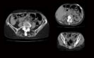
The use of contrast enhanced (CE) ultrasound for patients with abdominal trauma prior to computed tomography imaging has a higher diagnostic accuracy in comparison to conventional ultrasound according to the results of a systematic review and meta-analysis by researchers from the Brookdale University Hospital and Medical Center, New York, US.
Undertaking an extended Focused Assessment with Sonography in Trauma (eFAST) is commonly performed as part of the initial assessment of patients who have experienced trauma. Moreover, a systematic review has shown how eFAST serves as a useful bedside tool which is able to rule-in pneumothorax, pericardial effusion as well as intra-abdominal free fluid within a trauma setting but is less useful as a rule-out tool. The diagnostic capability of ultrasound can be improved with the use of a contrast media. In fact, contrast enhanced ultrasound in children has been found to have a comparable performance to both CT and MRI with a very high degree of specificity and hence has the potential to reduce irradiation exposure in paediatric patients. It has also been shown that contrast enhanced ultrasound seems to be both safe and accurate for the identification of abdominal solid organ injuries in children who have experienced a blunt abdominal trauma. Nevertheless, the comparative accuracy of CE ultrasound has not been directly compared to eFAST during the initial trauma assessment and was the subject of the review by the US researchers. The team focused on the use of both ultrasound methods for the initial assessment of patients with abdominal trauma before CT imaging. Paired pooled sensitivity and specificity were used to assess the relative merits of both approaches.
Contrast enhanced ultrasound diagnostic value in abdominal trauma
Following a literature search, a total of 10 eligible studies with 1,359 patients and 30 pairwise comparisons were included in the analysis.
Overall, the paired, pooled sensitivity for CE ultrasound was 0.933 (95% CI 0.917 – 0.948) compared to 0.559 (95% CI 0.527 – 0.591) for conventional ultrasound (p < 0.001). The pooled specificity was also significantly higher (0.995 vs 0.975, p < 0.001). In fact, in sub-group analysis, CE ultrasound was superior to conventional ultrasound for all other areas including the liver, kidneys and adrenals, spleen and for the presence of active bleeding.
Based on these findings, the authors concluded that CE ultrasound was a superior method for differentiating abdominal trauma injuries when used as an initial means of assessment in emergency departments.
Citation
Sutarjono B et al. Is it time to re-think FAST? A systematic review and meta-analysis of Contrast-Enhanced Ultrasound (CEUS) and conventional ultrasound for initial assessment of abdominal trauma. BMC Emerg Med 2023
21st October 2022
The use of a convolutional neural network (CNN) deep learning model significantly improved the diagnostic accuracy of orthopaedic surgeons’ detection of fractures as well as the time taken to make the diagnosis according to a study by Japanese researchers.
One of the main causes of diagnostic errors in the emergency department is the failure to correctly interpret radiographs and that the majority of diagnoses missed on radiographs are fractures. Moreover, in patients with multiple traumas, whole body CT scans are generally the preferred imaging modality, but a study has suggested that on a per-scan basis, there were 12.9% of missed injuries, of which 2.5% were clinically significant. The use of a CNN model for automatic detection of rib fractures detection could assist radiologists in improving diagnostic efficiency, reducing diagnosis time and radiologists’ workload. With some evidence that a CNN model can detect individual fractures, there remains uncertainty over whether such models could be of assistance in reducing missed fractures in a wide range of sites such as the spine, ribs and pelvis.
For the present study, the Japanese team examined whether a fracture detection algorithm could be of assistance to orthopaedic surgeons and compared their diagnostic accuracy with and without the CNN model. The team retrospectively reviewed patients who were diagnosed with either a pelvic, rib or spine fracture and who had undergone a CT scan within 7 days of their trauma. The CNN model was trained, and the performance assessed in terms of sensitivity, precision and the F1-score (which is a measure of the model’s accuracy).
CNN model and orthopaedic surgeon’s diagnostic performance
The training and validation set included 181 patients with a mean age of 54.3 (35.3% female). The testing set included 19 patients (mean age 51.2, 31.5% female) with 2,447 CT images and an average of 129 images per patients. The prevalence of pelvic, rib and spine fractures among the images were 5.8%, 3.6% and 5.5% respectively.
The CNN model itself had an overall mean sensitivity of 78.6%, a precision of 64.8% and an F1-score of 71.1% and also high for the individual fracture regions, e.g., pelvic (83.9%), rib (71.3%) and spine (78%).
Three orthopaedic surgeons with 3, 3 and years of experience respectively, reviewed the CT scans both with and without assistance from the CNN model.
For the detection of fractures of a whole-body CT scan, the mean sensitivity for the first surgeon was 69.7% alone but increased to 82.9% with the CNN model (p < 0.0001) and there were similar and significant differences for the other two surgeons. The model also improved the diagnostic accuracy for each of the three scanned areas.
When the researchers examined the time to diagnose the fracture, i.e., based on reading and interpreting the scan, the first surgeon diagnosed a fracture in 278.4 seconds, and this reduced to 162.3 seconds with assistance from the CNN model (p < 0.0001). Once again, there were significantly shorter diagnostic times for the other two surgeons.
The authors concluded that while their CNN model provided a good sensitivity for the detection of pelvic, rib and spinal fractures, when used by the orthopaedic surgeons their sensitivity improved as did the time to make a fracture diagnosis.
Citation
Inoue T et al. Automated fracture screening using an object detection algorithm on whole-body trauma computed tomography Sci Rep 2022
20th July 2021
The use of mitigation strategies for COVID-19 such as social distancing and the wearing of face-masks have helped to minimise the spread of the virus. In addition, the use of an effective Test-Trace-Isolate (TTI) strategy, involving the testing of symptomatic cases and tracing their contacts, is crucial for the detection of the virus. The current gold standard test is the polymerase chain reaction (PCR) although this testing modality can only be undertaken within a hospital laboratory and is therefore expensive for population-wide testing. While the testing process itself takes several hours, there are additional time constraints and which slow the overall process, e.g., transporting of samples to the laboratory testing site, presence of a sufficient number of trained staff and high sample volumes. An alternative is the use of lateral flow tests and these have been recognised as being able to increase testing capacity.
Lateral flow tests are are much cheaper than PCR tests and can be produced in large quantities and deliver results on site and within 15 to 30 minutes. Whereas PCR tests involve amplification of the nucleic acid sample, lateral flow tests rely on the detection of a viral antigen in the patient’s sample and are therefore deemed to be of lower sensitivity. However, there have been no comparative studies of outcomes associated with the use of PCR and antigen testing in the same patient cohort and this led a team from the Centre for Primary Care, Wolfson Institute of Population Health, Queen Mary University, London, to assess the diagnostic accuracy of both methods in the same patients. The study was undertaken among a network of general practitioners in Austria and included patients who self-reported mild to moderate flu-like illness (e.g., cough, fever, runny nose etc). Such individuals received a same-day appointment and given an antigen test and also given a PCR test.
Findings
Based on lateral flow tests for 2562 patients, 1027 who tested positive, were also given a PCR test and of whom, 826 (79.7%) tested positive. From this cohort of 826, 788 had also tested positive on the antigen test. The authors then calculated that overall sensitivity of the lateral flow tests to be 95.4%, with a specificity of 89.1%. In addition, positive lateral flow and PCR tests were correlated (r = 0.968).
The authors discussed how these findings indicated that using antigen tests for patients with mild to moderate symptoms, allows for a reliable and accurate detection of COVID-19 and which is comparable to PCR. They concluded that implementation of lateral flow tests should be accompanied by standardised training for operators, quality assurance of testing and a coordinated approach to services.
Citation
Leber W et al. Comparing the diagnostic accuracy of point-of-care lateral flow antigen testing for SARS-CoV-2 with RT-PCR in primary care (REAP-2). EClinicalMedicine 2021
IMPORTANT LINKS
MORE FROM COGORA
© Cogora 2025
Cogora Limited. 1 Giltspur Street, London EC1A 9DD
Registered in the United Kingdom. Reg. No. 2147432