This website is intended for healthcare professionals only.
Take a look at a selection of our recent media coverage:
14th November 2022
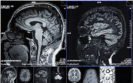
Assessment of S100B levels in low-risk head injury patients seen in an emergency department is a potential diagnostic marker to rule-out serious intracranial pathology that reduces the need for a CT-head scan in more than a quarter of cases according to a study by New Zealand researchers.
In a 2016 study it was estimated that globally, there were 27·08 million new cases of traumatic brain injury (TBI) caused primarily by falls and road injuries. Moreover, assessing patients with a head injury in an emergency department (ED) takes time with and has been estimated at 6.6 hours with the time related to head CT responsible for about half of the total length of stay.
Despite increased availability of CT scans and its potential value as a diagnostic tool, over 90% of head CT scans in those referred from ED to exclude a significant intracranial injury have been found to be normal prompting the search for alternative assessment tools. One potential biomarker protein is S100B, which has been described as a useful neuro-biochemical marker of brain damage such as in circulatory arrest, stroke and traumatic brain injury.
However, a recent systematic review concluded that the diagnostic accuracy of single biomarkers as a rule out for significant intracranial injury seen on CT head scans in ED patients with TBI is low. Nevertheless, the authors of the review added that S100B is the only single biomarker with a validated clinical platform making it the biomarker of choice. Despite this, there is little evidence on the diagnostic performance of S100B, particularly in low-risk head injury patients, where it is likely to be of greatest value in reducing the need for a CT-head scan.
In the present study, the New Zealand team examined the diagnostic value of the biomarker for excluding significant intracranial pathology in patients who presented at an ED within 6 and 24 hours of their head injury. Included patients were those able to give consent and without signs of post-traumatic amnesia and blood samples were taken to measure S100B levels.
All patients were assessed and sent for a CT-head scan when deemed clinically appropriate and categorised as CT-positive (i.e., needing a scan) or CT-negative. The primary outcome was the diagnostic accuracy of S100B within 6 hours after a head injury.
S100B diagnostic value in low-risk head injury
A total of 265 patients of whom, 133 with a median age of 69.5 years (35.8% female) presented within 6 hours of their head injury were included in the study.
Among those presenting within 6 hours, the median S100B level in those who were CT-positive was 0.36 and compared to 0.15 in the CT-negative group (p < 0.01). The sensitivity of S100B in the 6-hour group was 93.8% and the specificity of 30.8%.
The authors calculated that using a clinically validated cut-off threshold for S100B of 0.1 μg/mL, if patients with levels below this threshold did not have a CT-head scan, the use of scans would be reduced by 27.1%. Similarly, for those attending within 24 hours of their head injury and using the same threshold, would lead to a 37.4% reduction in CT scans. In fact, the authors also determined that the risk of missing a significant injury with this approach would result in missing only 0.75% of significant injuries within 6 hours or 2.3% within 24 hours.
They concluded measurement of S100B performed well as a diagnostic aid to exclude significant intracranial injury in low-risk patients with a head injury.
Citation
Rogan A et al. Diagnostic performance of S100B as a rule-out test for intracranial pathology in head-injured patients presenting to the emergency department who meet NICE Head Injury Guideline criteria for CT-head scan Emerg Med 2022
21st October 2022
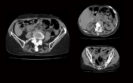
The use of a convolutional neural network (CNN) deep learning model significantly improved the diagnostic accuracy of orthopaedic surgeons’ detection of fractures as well as the time taken to make the diagnosis according to a study by Japanese researchers.
One of the main causes of diagnostic errors in the emergency department is the failure to correctly interpret radiographs and that the majority of diagnoses missed on radiographs are fractures. Moreover, in patients with multiple traumas, whole body CT scans are generally the preferred imaging modality, but a study has suggested that on a per-scan basis, there were 12.9% of missed injuries, of which 2.5% were clinically significant. The use of a CNN model for automatic detection of rib fractures detection could assist radiologists in improving diagnostic efficiency, reducing diagnosis time and radiologists’ workload. With some evidence that a CNN model can detect individual fractures, there remains uncertainty over whether such models could be of assistance in reducing missed fractures in a wide range of sites such as the spine, ribs and pelvis.
For the present study, the Japanese team examined whether a fracture detection algorithm could be of assistance to orthopaedic surgeons and compared their diagnostic accuracy with and without the CNN model. The team retrospectively reviewed patients who were diagnosed with either a pelvic, rib or spine fracture and who had undergone a CT scan within 7 days of their trauma. The CNN model was trained, and the performance assessed in terms of sensitivity, precision and the F1-score (which is a measure of the model’s accuracy).
CNN model and orthopaedic surgeon’s diagnostic performance
The training and validation set included 181 patients with a mean age of 54.3 (35.3% female). The testing set included 19 patients (mean age 51.2, 31.5% female) with 2,447 CT images and an average of 129 images per patients. The prevalence of pelvic, rib and spine fractures among the images were 5.8%, 3.6% and 5.5% respectively.
The CNN model itself had an overall mean sensitivity of 78.6%, a precision of 64.8% and an F1-score of 71.1% and also high for the individual fracture regions, e.g., pelvic (83.9%), rib (71.3%) and spine (78%).
Three orthopaedic surgeons with 3, 3 and years of experience respectively, reviewed the CT scans both with and without assistance from the CNN model.
For the detection of fractures of a whole-body CT scan, the mean sensitivity for the first surgeon was 69.7% alone but increased to 82.9% with the CNN model (p < 0.0001) and there were similar and significant differences for the other two surgeons. The model also improved the diagnostic accuracy for each of the three scanned areas.
When the researchers examined the time to diagnose the fracture, i.e., based on reading and interpreting the scan, the first surgeon diagnosed a fracture in 278.4 seconds, and this reduced to 162.3 seconds with assistance from the CNN model (p < 0.0001). Once again, there were significantly shorter diagnostic times for the other two surgeons.
The authors concluded that while their CNN model provided a good sensitivity for the detection of pelvic, rib and spinal fractures, when used by the orthopaedic surgeons their sensitivity improved as did the time to make a fracture diagnosis.
Citation
Inoue T et al. Automated fracture screening using an object detection algorithm on whole-body trauma computed tomography Sci Rep 2022
21st September 2022
A deep learning-based tool has been shown to accurately detect pancreatic cancers that are less than 2 cm and which can often be missed during an abdominal CT scan according to the findings of a retrospective study by Taiwanese researchers.
Pancreatic cancer has a poor prognosis and is the 12th most common cancer worldwide and in 2020 there were more than 495,000 new cases and an estimated 466003 global deaths. However, 5-year survival is poor and data for the UK suggests that only 7.3%)of people diagnosed with the cancer in England survive for five years or more.
The clinical diagnosis of pancreatic cancer is challenging as patients often present with non-specific symptoms with nearly a third of patients clinically misdiagnosed. Imaging has a crucial role to play in diagnosis though one retrospective analysis of different imaging modalities revealed that 62% of cases were missed and 46% misinterpreted, with 42% of cases missed because the tumour was less than 2 cm.
Previous research using a deep learning-based convolutional neural network, showed that the technology could accurately distinguish pancreatic cancer on computed tomography (CT) with acceptable generalisability to images of patients from various races and ethnicities.
However, in that study, segmentation of the pancreas, i.e. identifying that the region on a CT scan which actually is the pancreas, was performed manually by radiologists. But would it be possible for a deep learning-based tool to enable segmentation and to detect the presence of pancreatic cancer?
This was the question addressed in the current study by the Taiwanese team. They used contrast-enhanced CT collected from patients who had been diagnosed with pancreatic cancer and compared these with CT scans of non-cancer, control patients.
The deep learning-based tool was initially trained and validated on samples with and without cancer and then tested in a real-world set of CT scans and its performance assessed based sensitivity, specificity and accuracy.
Deep learning tool and prediction of small pancreatic tumours
A total of 546 patients with a mean age of 65 years (46% female) who had pancreatic cancer with a mean tumour size of 2.9 cm and 733 control patients were used in the training, validation and test set.
In a nationwide test set that included 669 cancer patients and 804 controls, the deep learning-based tool distinguished between CT malignant and control samples with a sensitivity of 89.7% (95% CI 87.1 – 91.9) and a specificity of 92.8% (95% CI 90.8 – 94.5) and an accuracy of 91.4%.
When comparing the tool with radiologists, the corresponding sensitivities for the local test set (109 cancer and 147 control patients) were 90.2% and 96.1% for the tool and radiologists respectively and this difference was not significant (p = 0.11).
The tool had a sensitivity of 87.5% (95% CI 67.6 – 97.3) for a malignancy which was smaller than 2 cm in the local test set although this was slightly lower (74.7%) in the nationwide test set.
The authors concluded that their tool may be of value as a supplement for radiologists to enhance detection of pancreatic cancer although further work was needed to examine the generalisability of the findings of other populations.
Citation
Chen PT et al. Pancreatic Cancer Detection on CT Scans with Deep Learning: A Nationwide Population-based Study Radiology 2022
27th July 2022
Combining an additional magnetic resonance imaging (MRI) and computed tomography (CT) scan in patients presenting with an acute ischaemic stroke, does not lead to better patient outcomes at both discharge and 12 months later, according to the findings from a retrospective study by US researchers.
Data from 2017 show that there were 1.12 million incident strokes in the European Union, 9.53 million stroke survivors, 0.46 million deaths, and 7.06 million disability-adjusted life years lost because of stroke. Patients who present with an acute ischaemic stroke (AIS) are normally initially evaluated with a CT scan to exclude haemorrhage.
Moreover, according the the American Heart Association/American Stroke Association guidelines, in patients who are suspected of having ischaemic stroke, if CT or MRI does not demonstrate symptomatic cerebral infarct, follow-up CT or MRI of the brain is reasonable to confirm diagnosis. In support of this recommendation, there are already some data to indicate that the use of MRI scanning in patients with an AIS is associated with substantial decrease in the rates of inpatient mortality and complications.
However, whether combining a CT scan with an additional MRI improves outcomes for patients is less clear. To date, only a single study has examined the effect of a CT scan and an additional MRI scan on patient outcomes, concluding that the long-term (one-year) patient outcomes may not be influenced by either imaging strategy.
In trying to better understand the value of a strategy that combined a CT scan with an additional MRI scan, in the present study, the researchers undertook a retrospective, propensity score matched study of the clinical outcomes at both discharge and one year later, for patients hospitalised with an AIS.
For the outcomes of interest, the researchers used either death or dependence at discharge, based on the Modified Rankin scale (mRS) which assesses disability in patients who have suffered a stroke. The scale ranges from 0 (no disability) through 5 (requiring constant care) to 6 (death) and the researchers set a non-inferiority margin of -7.5%, for the percentage of patients discharged with a mRS score of 3 to 6. For death, the researchers set a relative risk, RR of 0.725 as the margin for non-inferiority.
Additional MRI and clinical outcomes
A total of 246 patients (123 with and without an MRI scan) and a median age of 68 years (53% male) were included in the study. Among those who had an additional MRI scan, this was ordered by the attending vascular neurologists in 42.3% of cases, 33.3% by emergency physicians and 24.4% by nurse practitioners. In 111 cases of the 123 MRIs ordered, there was no specific indication other than either a stroke or neurological symptoms.
For the primary endpoint of a mRS score 3 – 6 at discharge, there was no significant difference between the two groups (relative risk, RR = 1.50, 95% CI 0.86 – 1.05, p = 0.44), with an absolute difference of 5.7%, thus meeting the -7.5% difference criteria for inferiority.
Stoke or death at one year after discharge occurred in more patients receiving the additional MRI compared to those who only had a CT scan (19.5% vs 12.5%), giving a RR of 1.14 (95% CI 0.86 – 1.50) thus again meeting the RR of 0.725 criteria for non-inferiority.
The authors concluded that the value of an additional MRI to a CT scan in patients with AIS should not be presumed and called for future studies to identify which patients hospitalised with an AIS may benefit from an MRI scan.
Citation
Frade HC et al. Comparison of Outcomes of Ischemic Stroke Initially Imaged With Cranial Computed Tomography Alone vs Computed Tomography Plus Magnetic Resonance Imaging JAMA Netw Open 2022
4th July 2022
The introduction of an alert system to provide clinical decision support in emergency departments on whether to perform a computed tomography (CT) scan on children with head trauma, has resulted in more appropriate use of such scans. This was the main conclusion of a study by a team from Utah, US.
Traumatic brain injury (TBI) is an important public health problem and data produced by the CDC in the US reveal how there are approximately half a million emergency department visits for TBI every year by children aged 0 to 14 years.
An important concern for clinicians is to identify whether a child has a clinically important TBI and a CT scan represents an excellent imaging modality for the identification of an intracranial injury. However, not all children with a head injury require a CT scan and clinical decision rules can help standardise and improve the use of CT for children with minor head injury.
For the present study, the US wanted to explore whether the introduction of an alert system at the point when clinicians were considering the use of a CT scan, could provide enhanced support and therefore make more appropriate use of these scans.
The alert system made use of the Pediatric Emergency Care Applied Research Network (PECARN) head trauma clinical decision rules, which provided risk stratification to help reduce diagnostic uncertainty and the need for unnecessary CT scans. Prior to the decision-making for the need to perform a CT scan, clinicians were presented with a pop-up’ alert, which provided a risk assessment.
In trying to assess the value of introducing the alert system, the researchers considered guideline adherence, which they considered to be not performing a CT scan when the alert system identified the patient as being at low risk.
For the study, all sites provided data on the level of CT scans before and after introduction of the intervention for comparative purposes.
Alert system and change in CT scanning
A total of 12, 670 paediatric minor head trauma encounters were included in the analysis. The proportion of guideline-adherent encounters increased from 94.8% in the control period to 99.4% during the time when the intervention was implemented. Furthermore, the proportion of CT scans performed reduced from 38.6% in the control period to 29.8% after implementation of the intervention.
The authors calculated that the odds of an encounter being guideline-adherent was 1.12 (95% CI 1.03 – 1.22) or approximately 10% higher during the intervention compared to the control time period. Moreover, when using a pre-post comparison, guideline adherence remained significantly higher than the control period (odds ratio = 5.33, 95% CI 3.75 – 7.59).
The authors concluded that the implementation of the alert system led to sustained improvements in adherence to guidelines on the need for a CT scan and a modest reduction in scans among low risk patients.
Citation
Knighton AJ et al. Improving Head CT Scan Decisions for Pediatric Minor Head Trauma in General Emergency Departments: A Pragmatic Implementation Study Ann Emerg Med 2022
27th June 2022
The presence of incidental findings (IF) occurs in roughly a third of all computed tomography (CT) scans undertaken with emergency departments according to the findings of a systematic review by a group of US researchers.
The term ‘incidentaloma’ refer to an incidentally discovered mass or lesion, detected using imaging and which was performed for an unrelated reason. Such incidental findings are not uncommon, especially among scans for trauma patients with one study revealing how the findings were present in 15% of trauma CT scans.
Whilst the presence of IF do not affect or alter the emergency department clinician’s current diagnostic work-up, it is important that these observations are communicated to patients so as to ensure that where necessary, appropriate further tests and follow-up are instigated. However, in non-trauma patients, little is known about the level of incidental findings among CT scans undertaken within emergency departments.
For the present study, the US team sought to estimate the prevalence of radiologic IF among patients visiting an emergency department and who underwent a CT scan. A secondary aim was look at how hospitals managed and stratified the risks associated with abnormal findings.
Undertaking a comprehensive literature search, the authors looked for studies including terms such as ‘incidentaloma’ without a restriction on the type of study design and which included those that were retrospective, cross-sectional or prospective in nature. The primary outcome of the systematic review was the prevalence of IF on a CT scan.
Prevalence of incidental findings on CT scans
A total of 69 studies representing 147,763 emergency department encounters or radiology reports, with a median patient sample size of 882, were included in the analysis.
The majority of studies were cross-sectional in design (82.6%) with the remainder comprising cohort (7.2%) and those with a pre- and post-interventional design. Just over half of the studies (50.7%) were in trauma patients and 63.8% of studies included some form a risk stratification of IF.
The pooled prevalence of any incidental finding on CT scan was 31.3% (95% CI 24.4% – 39.1%) although there was marked evidence of heterogeneity in the studies. The highest prevalence of IF occurred in patients having a CT scan because of chest pain (36.6%), followed by trauma (34.7%).
In a total of five studies, all based in trauma centres, there were structural interventions designed to improve the recognition and notification or follow-up of patients identified with an IF on a CT scan. In one such study, for example, following implementation of the strategy to manage IF, patient notification increased from 17.7% to 32.4%.
The researchers also discovered that documentation of IF in the patient’s discharge notes was present in only 20.1% to 47.2% of cases.
In their conclusion, the authors identified the need to establish a comprehensive classification system and standard-based approach to help clinicians when faced with an IF. They also called for more flexible care co-ordination programs to ensure timely follow-up, clear documentation in medical records and which could easily be implemented within a busy emergency department.
Citation
Evans CS et al. Incidental Radiology Findings on Computed Tomography Studies in Emergency Department Patients: A Systematic Review and Meta-Analysis Ann Emerg Med 2022
20th September 2021
According to Cancer Research UK, there are around 47,800 new lung cancer cases each year and approximately 35,100 deaths, which equates to 96 deaths every day. Furthermore, Cancer Research UK estimates that 79% of lung cancer cases in the UK are preventable with 72% caused by smoking. With such a high incidence of not only cases, but more importantly, preventable cases, there is an urgent need for effective screening methods, especially among individuals who are deemed at high risk such as smokers. In a 1999 study, a low computed-tomography (CT scan) was shown to greatly improve the likelihood of detecting small, non-calcified nodules and hence lung cancer, at an earlier and hence more curable stage. Moreover, subsequent studies have also demonstrated a reduction in lung cancer mortality among those undergoing a low dose CT scan.
With the value of CT screening already firmly established, a UK-based team have published their own findings of a trial comparing the effect of a low dose CT scan compared to usual care, in high-risk patients. The UK lung cancer screening (UKLS) trial, randomised patients to low dose CT screening or usual care, i.e., with no CT scan and was undertaken at two thoracic hospitals in the UK. Eligible patients, aged 50 to 75 years, were those deemed to be at a high risk of developing lung cancer over the next 5 years defined by a risk score of at least 4.5% based on the Liverpool Lung Project risk model (LLPv2). This model includes several possible risk factors such as gender, age, smoking status, smoking duration, family history of lung cancer. Included patients were then randomised to the intervention group (CT scan) or usual care although given the nature of the intervention, blinding was not possible. The primary outcome was mortality due to lung cancer, defined as a death during the follow-up period where lung cancer was listed as an underlying cause. In an effort to provide further evidence, the researchers also undertook a meta-analysis of other recent trials and included their own data, to get a more robust estimate of the benefits of CT scanning.
Findings
A total of 1987 and 1981 individuals were randomised to the CT scan and control arm respectively and followed for a median of 7.3 years. The median age at consent was 68 years (25% female) and among the CT scan group, 38% were current smokers, of whom, 93% had smoked for more than 20 years. During the follow-up period, 76 lung cancers were detected, 30 in the CT scan arm and 46 in the control arm although this difference was not significant (relative risk, RR = 0.65, 96% CI 0.41 – 1.02, p = 0.062). Furthermore, there were no significant differences between the sexes. In addition, there were 512 deaths from any cause and again there was no significant difference between the groups (p = 0.315).
When these results were added to a meta-analysis of 9 randomised, controlled trials, low dose CT scan screening was associated with a 16% relative reduction in lung cancer mortality compared with no screening (RR = 0.84, 95% CI 0.76 – 0.92).
The authors concluded that while their trial had not demonstrated a statistically significant reduction in lung cancer mortality, when their data was combined with other studies, the pooled estimate was significant and provided further support for lung cancer screening via a low dose CT scan.
Citation
Field JK et al. Lung cancer mortality reduction by LDCT screening: UKLS randomised trial results and international meta-analysis. Lancet Regional health Europe 2021
IMPORTANT LINKS
MORE FROM COGORA
© Cogora 2025
Cogora Limited. 1 Giltspur Street, London EC1A 9DD
Registered in the United Kingdom. Reg. No. 2147432