This website is intended for healthcare professionals only.
Take a look at a selection of our recent media coverage:
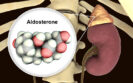
21st January 2023
Metomidate used as a PET radiotracer and in combination with high resolution computed tomography (CT) has been shown to be non-inferior to adrenal vein sampling (AVS), as a non-invasive means of detecting primary aldosteronism according to a study by a team of UK researchers.
Primary aldosteronism (PA) has been shown to be responsible for 5.9% of cases of hypertension and this figure increased to 11.8% in patients with stage 3 hypertension. PA can be surgically corrected when caused by unilateral aldosterone hyper-secretion for which the usual cause is an aldosterone-producing adenoma. Moreover, hypertension due to PA has a worse prognosis compared with blood pressure-matched essential hypertension. The use of AVS is recognised as the most reliable means of identifying whether aldosterone production is uni- or bilateral. Nevertheless, an alternative is the use of imaging modality, such as metomidate positron emission tomography computed tomography (MTO) and which, in one small study has been shown to both a sensitive and specific alternative to adrenal vein sampling. Nevertheless, with a limited evidence base to demonstrate the value of this imaging modality, in the present study, researchers set out to compare the accuracy of MTO and AVS at predicting the outcome following adrenalectomy in patients with PA and ultimately resolution of hypertension in these patients. Individuals with confirmed PA underwent both MTO and AVS and were commenced on spironolactone 50mg but which was increased to 100mg after two weeks. Both techniques were used to assess the probability of unilateral PA and where this was high, unilateral adrenalectomy was recommended, or medical management where it was not detected.
Metomidate scanning and the outcome following adrenalectomy
A total of 128 patients with a median age of 52 years (68% male) were included in the study.
The use of MTO graded 52% of patients with a high probability of unilateral PA compared with 45% following AVS, although overall, 67% of the entire cohort were scored as having a high probability of unilateral PA.
Following surgery, the accuracy of MTO at predicting clinical success was 65.4% compared to 61.5% for AVS. These differences did not reach the predefined inferiority statistical margin; in other words, MTO was not inferior to AVS. Only 23 of 78 patients undergoing surgery achieved blood pressure readings of below 135/85mmHg, although 12 individuals were able to stop antihypertensive treatment.
There were a total of 24 serious adverse events although none of these were considered to be related to the procedures, and 22 fully resolved.
The authors concluded that MTO was an effective non-invasive means to diagnose unilateral PA and could be used as an alternative to AVS.
Citation
Wu X et al. [11C]metomidate PET-CT versus adrenal vein sampling for diagnosing surgically curable primary aldosteronism: a prospective, within-patient trial. Nat Med 2023
4th October 2021
Acute chest pain is a common reason for attendance at an emergency department, accounting for approximately 10% of non-injury-related visits. While chest pain can arise from non-cardiac causes, all those presenting with chest pain will be assessed for acute coronary syndrome (ACS). Prompt treatment is required for patients with obvious clinical signs and symptoms of ACS whereas those deemed to be at either a low or intermediate risk initially undergo observation and further testing only if ACS is suspected. One strategic approach to the assessment of low risk ACS patients, is early computed tomography (CT) coronary angiography (CTCA). This technique has been increasingly used to assess patients with stable chest pain because it has high sensitivity and specificity for the detection of coronary heart disease. Within an emergency care department, CTCA therefore allows for a rapid evaluation of patients presenting with acute chest pain. In fact, one systematic review concluded that the use of CTCA is associated with a reduced length of hospital stay compared to usual care. Furthermore, a 2018 study examining the use of CTCA in patients with stable chest pain, found that over a 5-year period, there was a significantly lower rate of death from coronary heart disease or non-fatal myocardial infarction compared to standard care. However, while studies have focused on patients at low risk of ACS, whether early CTCA is of value in those patients deemed to be at an intermediate risk of ACS is less clear.
In trying to assess the value of early CTCA in patients presenting with acute chest pain and at an intermediate level of risk for acute coronary syndrome, researchers from Edinburgh University, established the RAPID CTCA study. The trial enrolled adult patients with suspected or a provisional diagnosis, of acute coronary syndrome and prior coronary heart disease. These individuals where then randomised 1:1 after admission to hospital to early CT coronary angiography with standard care or standard care alone. The primary outcome was the time to the first event of all-cause death or a subsequent non-fatal type 1 (spontaneous) or type 4b (related to stent thrombosis) myocardial infarction at one-year. Secondary outcomes included the cause of death and subsequent myocardial infarction.
Findings
The study recruited and randomised 1748 patients with a mean age of 61.6 years (64% men), of whom 877 received early CTCA. Overall, 89% of patients had chest pain as their primary complaint, 34% had existing coronary heart disease, 57% raised cardiac troponin levels and 61% an abnormal ECG. The primary outcome all cause death or non-fatal myocardial infarction (both types) occurred in 5.8% of those in the early CTCA group and 6.1% of those assigned to usual care (adjusted hazard ratio, aHR = 0.91, 95% CI 0.62 – 1.35, p = 0.65). Furthermore, there were no significant differences in any of the secondary outcomes. The need for invasive coronary angiography occurred in 54% of those in the CTCA group and 60.8% in the usual care group (aHR = 0.81, 95% CI 0.72 – 0.92, p = 0.001).
The authors concluded that early CTCA for patients with an intermediate risk of ACS did not alter overall coronary therapeutic interventions or one year clinical outcomes but did reduce the need for invasive angiography.
Citation
Gray AJ et al. Early computed tomography coronary angiography in patients with suspected acute coronary syndrome: randomised controlled trial. BMJ 2021
20th September 2021
A squamous cell carcinoma on the head or neck is the sixth most common cancer globally, with around 890,000 new cases and 450,000 deaths in 2018. The main form of treatment is curative radiotherapy and in patients with locoregionally advanced cancers, prior scanning with fluorodeoxyglucose positron emission tomography and computed tomography (PET-CT) has been shown to have good diagnostic performance for the detection of regional nodal metastasis. However, where there is a delay between radiotherapy and the initial PET-CT scan, does this impact on radiotherapy planning and might it be necessary to perform a second scan prior to radiotherapy? This was the question posed by researchers from the Department of Radiation Oncology, Inselspital, Bern University Hospital, Bern, Switzerland. The team performed a retrospective analysis of patients with advanced head or neck squamous cell carcinoma and who had received two PET-CT scans prior to radiotherapy, to determine whether the second scan led to any modifications to radiotherapy treatment. The team looked for changes in the primary tumour, lymphatic spread and the presence of distant metastases between the two scans. They categorised any changes as minor if there were modifications to the RT plans such as dose changes and major where treatment moved from curative to palliative or the addition of induction chemotherapy, a switch to surgery or any additional diagnostic work-up that led to postponement or cancellation of treatment.
Findings
There were 32 newly diagnosed patients with locoregionally advanced squamous cell cancer with a median age of 64 years (34% female). The median interval between the initial scan for staging assessment and the second scan was 42.5 days. Just over half (53%) of patients had a grade 2 and 41% a grade 3 tumour. Fortunately, a major treatment change occurred in only 1 patient although nodal upstaging occurred in 10% (3/29) of patients. Minor treatment changes were required in 52% (16/31) of patients with new lymph node metastases detected in all 16 patients and in 6 cases, there was evidence of progression of the primary tumour size.
In discussing their findings, the authors noted that despite an initial PET-CT scan to assess tumour staging, a second scan identified the need for minor changes in just over half of all patients. Based on these findings, they called for the potential benefits of a second scan to be further investigated and validated. They also noted that the practice of undertaking a second scan of patients where the delay was more than four weeks has become the established practice at their hospital.
Citation
Elicin O et al. Impact of pre-treatment second look 18FDG-PET/CT on stage and treatment changes in head and neck cancer. Clin Trans Radiat Oncol 2021
IMPORTANT LINKS
MORE FROM COGORA
© Cogora 2025
Cogora Limited. 1 Giltspur Street, London EC1A 9DD
Registered in the United Kingdom. Reg. No. 2147432