This website is intended for healthcare professionals only.
Take a look at a selection of our recent media coverage:
10th December 2021
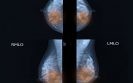
The presence of subclinical axillary lymphadenopathy (sLAD) on mammography was significantly more likely among women who had received a COVID-19 vaccination, indicating that they should either delay screening or schedule visits before vaccination. This was the finding of a retrospective study by a team from the Baylor University Medical Center, Dallas, USA.
The term lymphadenopathy refers to lymph nodes that are abnormal in size and axillary lymphadenopathy, describes changes in the size and consistency of lymph nodes in the armpit. There are several potential causes of unilateral axillary lymphadenopathy including breast cancer and which therefore warrants further diagnostic investigations if these changes are observed during screening. Although the presence of sLAD has been observed in women who have received a COVID-19 vaccination, what remains uncertain, is the proportion of vaccinated women that will develop these lesions after either the Pfizer or Moderna vaccines.
Based on case reports of sLAD in vaccinated women, for the present study, the authors investigated the prevalence of the condition in all women who had attended a breast screening programme since December 2020 until April 2021. Data from medical records included the woman’s vaccination status, the vaccine received and the presence or absence of sLAD on mammography. The researchers cross-referenced prior imaging for all women to ensure that there was no baseline evidence of an axillary lymphadenopathy and excluded women who had presented for diagnostic mammography for a palpable axillary lump or pain and those with a history of breast cancer.
Findings
In total, 1027 women with a mean age range of 56.4 to 63.7 years were included in the analysis. There were 158 who received the Moderna vaccine, 144 (Pfizer) and 725 who had not been vaccinated. From these 1027 women, 43 (4.2%) had an sLAD; 3.3% of those who had been vaccinated compared to 0.9% who were unvaccinated and this difference was statistically significant (p < 0.01).
When comparing the two vaccines, the incidence of unilateral axillary lymphadenopathy was higher among those who received the Pfizer vaccination (13.2% vs 9.5%, Pfizer vs Moderna). Among the 43 women with sLAD and who were recalled after screening, vaccine induced sLAD was more likely to have resolved when re-scanned an average of 46.5 days since their COVID-19 vaccination.
The authors concluded that women who have received a COVID-19 vaccination within 8 weeks of their screening mammogram, were significantly more likely to present with a subclinical axillary lymphadenopathy and that this could result in unnecessary further diagnostic workload. They suggested that providers should either delay screening mammograms by 8 weeks in patients who have been vaccinated or that screening should be undertaken before vaccination to avoid unnecessary follow-up.
Citation
Raj S et al. COVID-19 vaccine-associated subclinical axillary lymphadenopathy on screening mammogram Acad Radiol 2021
15th October 2021
The risk of breast cancer is increased among women with more dense breasts and the use of mammography can often miss cases in women with denser breasts. In a 2019 trial it was found that the use of supplementary breast MRI screening in women with dense breasts, lead to the diagnosis of significantly fewer interval cancers than mammography alone. However, screening programmes involve a huge number of women and many breast MRI scans of women with dense breasts show normal anatomical and physiological variation and therefore may not require radiological review.
A team from the Department of Radiology, University of Utrecht, therefore wondered if it was feasible to use an automated deep learning (DL) system for breast MRI screening to triage out normal scans without cancer to reduce the workload of radiologists. The team undertook a secondary analysis of data obtained from the prospective Dense Tissue and Early Breast Neoplasm Screening (DENSE) trial and the DL system was trained on left and right breasts separately and the results combined so that it was able to to differentiate between breasts with and without lesions. The performance of the DL system was assessed using the receiver operating characteristics (ROC) curves.
Findings
A total of 4581 breast MRI examinations of extremely dense breasts from 4581 women with a mean age of 54.3 years were included in the analysis. Of these 9162 breasts, 838 had at least one lesion, of which 77 were malignant. The area under the ROC curves in differentiating between a normal breast MRI and an examination with lesions was 0.83 (95% CI 0.80 – 0.85). The DL system considered that 90.7% (95% CI 86.7 – 94.7) of the MRI examinations with lesions were considered to be non-normal and would therefore be triaged to a radiologist review. In addition, the DL system dismissed 39.7% of the MRI examinations without lesions but did not miss any cases of malignant disease.
Commenting on their findings, the authors recognised a limitation in that their results were from the first round of the DENSE trial and that the number of lesions detected in subsequent screening rounds was smaller. Thus, they planned to further validate the performance of the model on data from subsequent rounds. The authors also suggested that future trials need to focus on demonstrating that the DL system is at least as effective as an expert radiologist at dismissing normal MRI examinations.
Citation
Verburg E et al. Deep Learning for Automated Triaging of 4581 Breast MRI Examinations from the DENSE Trial. Radiology 2021
IMPORTANT LINKS
MORE FROM COGORA
© Cogora 2025
Cogora Limited. 1 Giltspur Street, London EC1A 9DD
Registered in the United Kingdom. Reg. No. 2147432