This website is intended for healthcare professionals only.
Take a look at a selection of our recent media coverage:
20th January 2023
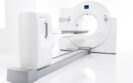
Using contrast-enhanced cone beam breast CT provides a more accurate assessment of residual tumour following neoadjuvant chemotherapy compared to magnetic resonance imaging (MRI) according to the findings of a comparative study by Chinese researchers.
Neoadjuvant chemotherapy is used prior to breast cancer surgery in patients with locally advanced breast cancer to reduce the size of the tumour. An assessment of the size of unresectable residual tumour is required since this is an important factor in the local recurrence of disease following breast conserving therapy. Radiological examination has an important role in the assessment of residual tumour and MRI has been shown to be more accurate that other imaging modalities for an evaluation of the response to treatment. Cone beam breast CT is a relatively novel technique that has shown promise for the early diagnosis of malignant breast cancer and can also differentiate between malignant and benign breast tissue.
However, to date, no studies have examined the accuracy of cone beam breast CT for the assessment of residual tumour following neoadjuvant chemotherapy. In the current study, the Chinese team compared cone beam breast CT and MRI for the assessment of tumour size following neoadjuvant chemotherapy and retrospectively compared the results of the two methods with those obtained with the findings from pathology. In addition, the researchers examined the predictive value of the two approaches for a pathological complete response. The level of agreement between the tumour size based on the two methods was compared to the findings on pathology and assessed using the intraclass correlation coefficient (ICC).
Cone beam breast CT and assessment of residual tumour
Data were available for 91 women with a median age of 45 years, the majority (73.6%) of whom were premenopausal.
When compared with pathology, there was good agreement for the cone beam breast CT (ICC = 0.64, 95% CI 0.35 – 0.78). In contrast, comparison with MRI was only moderate (ICC= 0.59, 95% CI 0.36 – 0.77). In subgroup analysis, the cone beam was also superior to MRI for residual ductal carcinoma in situ (p < 0.001).
For predictive purposes, the area under the receiver operating characteristics curve for predicting a pathological complete response were similar for both imaging modalities (AUC = 0.749 for cone beam and 0.733 for MRI, p > 0.05).
The authors concluded that cone beam breast CT was superior to MRI for an assessment of residual tumour following neoadjuvant chemotherapy and could therefore be seen as an alternative means of assessment.
Citation
Wang Y et al. Accuracy of Preoperative Contrast-enhanced Cone Beam Breast CT in Assessment of Residual Tumor after Neoadjuvant Chemotherapy: A Comparative Study with Breast MRI. Acad Radiol 2023
IMPORTANT LINKS
MORE FROM COGORA
© Cogora 2025
Cogora Limited. 1 Giltspur Street, London EC1A 9DD
Registered in the United Kingdom. Reg. No. 2147432