This website is intended for healthcare professionals only.
Take a look at a selection of our recent media coverage:
11th June 2024
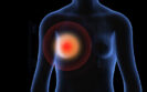
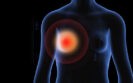
Consultant radiologist Dr Gerald Lip took an early interest in the use of artificial intelligence in medicine. He tells Steve Titmarsh how his team’s work in Aberdeen is unveiling the technology’s potential to augment clinicians’ workflows in breast cancer screening, which they hope will improve patient outcomes and help colleagues manage an ever-increasing demand for their services.
It was a master’s degree in health informatics at Trinity College Dublin that ignited Dr Gerald Lip’s interest in artificial intelligence and its applications in medicine. And while his medical career began in surgery, after a short time he concluded that the world of radiology was better suited to him and so he commenced this training at Aberdeen Royal Infirmary.
Here, he was fortunate to be guided by an excellent academic team, including Professor Fiona Gilbert, now at Cambridge University, who led a number of national trials in breast screening and imaging, and was recently appointed the first clinical radiology AI lead advisor at the Royal College of Radiologists. It was this experience that brought Dr Lip to his speciality in breast screening, and after two years as a consultant he was appointed clinical director of the North East Scotland Breast Screening Programme.
In 2018, the agency Innovate UK wanted to establish five centres of excellence for AI, and one of these was based in Scotland, encompassing Aberdeen and Glasgow. Here, a five-year programme called iCAIRD – the Industrial Centre for Artificial Intelligence Research in Digital Diagnostics – was established and, for Dr Lip, the opportunity to determine the potential of AI in breast screening was a huge draw.
Dr Lip’s involvement in the iCAIRD radiology collaboration began with a retrospective study using a three-year dataset (1 April 2016 to 31 March 2019) from the Scottish Breast Screening Service.
Published in the journal Radiology: Artificial Intelligence in 2023, the results highlight the possible value of an AI tool to augment the work of human scan readers for breast screening.
This was only the beginning. Dr Lip and his team recently completed a prospective trial to evaluate the AI tool in a clinical setting, using it in addition to two human readers of breast scans in work funded by the NHS Health and Social Care Award.
The work was part of a project named GEMINI (Grampians Evaluation of Mia in an Innovative breast screening Initiative) – a collaboration led by the NHS Grampian Innovation Hub; Kheiron Medical Technologies, which developed the AI tool; and the University of Aberdeen.
The project uses AI to evaluate anonymised patient images in a secure and trusted environment for data processing. ‘We have a very strong information governance team in the hospital so what happens is screening images are stripped of identifiable patient information and given a pseudonymised number before being sent to the cloud. The AI does its analysis and then it comes back and within the NHS its rejoined [with the patient data],’ Dr Lip explains.
Some early initial results from GEMINI were presented at the European Congress of Radiology in March 2024. These revealed that by using AI as a ‘safety net’ image reader, 11 women were found to have cancer who would likely otherwise not have been detected until their next routine breast screening. As a result, they were detected at an earlier stage when their cancers were smaller.
This discovery has significant implications for breast screening, patient outcomes and health system resource. For example, ‘if a cancer smaller than 15mm in size is detected, generally the chance of survival is 95%’, Dr Lip says. ‘[A] key part of screening is we want to find small things before they become big things, and AI is one of the tools that can help us find these small things.’
In that way, this AI tool can act as a safety net, which can have positive implications for patients and reduce the need for more complex surgery, chemotherapy and radiotherapy.
Dr Lip’s team is currently working on a paper looking into the health economics of AI. ‘Similar work with chest X-rays by researchers in Glasgow shows that costs rise in the first two years of implementing AI due to greater detection rates,’ he explains. ‘But later, as the technology is scaled up, cost savings result from early detection and the reduced need for more expensive interventions for more advanced disease states.’
The next stage of this research will be to look at a larger dataset: Dr Lip’s team are co-applicants for an National Institute for Health and Care Research grant for a trial to evaluate AI in a UK-wide breast screening programme.
A key development in the evolution of breast cancer screening came in around 2015/16 in the switch from film to digital imaging. Traditional film imaging does not lend itself to the use of AI tools because the scanned film images are poor quality compared with digital counterparts. That development, coupled with the simultaneous rise of AI, led to their obvious pairing to enhance clinicians’ work.
The technology used in medical imaging, for example, is not the same as the much-discussed large language models such as ChatGPT. Instead, the AI used in the GEMINI project is very much a tool that can be fine-tuned to suit a specific use – and even a particular population – to produce the best results.
Another future benefit that Dr Lip hopes will result from employing AI is the automation of screening normal mammograms, reducing clinician workload. ‘Currently two radiologists look at each scan,’ he explains. ‘In future, if the AI records a mammogram as normal and a human screener agrees, there may be no need for a second human to verify the result.’
Indeed, initial results from an evaluation of AI using data modelling predicted a 30% reduction in clinician workload. Dr Lip explains that in a real-world situation this could mean his centre, where they read 20,000 mammograms a year, would not need a second human to read around 5,000 mammograms. This would free up a significant amount of clinician time for other tasks such as biopsies or face-to-face time with patients, he says.
Given that the Royal College of Radiologists estimates a 29% shortage of radiologists, which will rise to 40% in five years without action, and with breast screening likely to be one of the most affected specialties due to rising breast cancer cases in women, the potential time saving afforded by using an AI tool could be significant.
What’s more, as breast screening in Scotland sits under a national Picture Archiving Communications System (PACS), Dr Lip says scaling the GEMINI project to cover the whole country could be achieved ‘quite easily’. This could also be the case for Northern Ireland, which runs a unified PACS programme, as well as in England’s emerging imaging networks.
For Dr Lip, a key message from his research so far is that AI should be seen as supplementary and not a replacement for clinicians’ expertise – an experienced human still needs to be in the loop.
Of course, another reason for human involvement is that the AI only analyses the data it is given so it would not automatically know about previous surgeries and biopsies, unless this data was inputted. It may also unnecessarily recommend call-backs based on lesions from previous surgery, for example, whereas a human investigator would have access to the patient history and be able to exclude such scans from being re-read.
Dr Lip says that another significant factor highlighted was the importance of effective monitoring. AI systems suffer from drift, which happens when the software, machine or population changes, and it means that the performance of the AI responses slowly degrade over time. Monitoring is therefore needed to ensure the performance remains as expected and required.
And then there’s bias, particularly in terms of different patient populations. It is important to ensure there is no bias for different ethnic groups, for example.
‘Bias is a hot word in the AI world to ensure that it’s not biased against one group,’ says Dr Lip. ‘If an AI is only trained on white women, are there are differences in the mammograms of white women compared to black women? We don’t have significant knowledge to that level and it’s a fast-moving space.’
Real-world data from a representative population that the AI tool is analysing needs to be used to try to reduce biased results.
Looking ahead, Dr Lip says ‘more than one AI tool may be used to assess breast scans’, and he is aware of this currently happening in other clinical areas. ‘For example, in stroke care, there is a tool trained on the black areas of scans and a second trained on the white parts, so using the two tools in conjunction could improve results,’ he explains.
Another area of research for Dr Lip is in so-called masking. If there is a lot of glandular tissue in a breast, it can mask tumours and AI seems to perform well in these cases, he says. Colleagues at the Karolinska Institute in Sweden have developed a masking tool and a density tool, which can be used along with the AI screening tool, and there is more work to be done in this area.
Genetics is also key to revolutionising breast cancer care. At the Aberdeen Centre for Health Data Science, in collaboration with Professor Lesley Anderson and her team, Dr Lip is undertaking multidisciplinary work in this field.
Using BioBank data in breast cancer, the aim is to advance treatment personalisation. In future, by combining scan data with genetic and biopsy information, for example, it may be possible to determine how often each woman should attend screenings to ensure cancers are detected as early as possible.
With all their published work and a host of novel projects in the pipeline, it’s clear that the work Dr Lip and his team in Aberdeen are doing with AI promises to help alleviate workforce pressures in the NHS, detect more cancers at an earlier stage and, ultimately, improve outcomes for patients.
23rd May 2024
Adopting a risk-stratified approach to breast cancer screening could help improve the benefit-to-harm ratio and the cost-effectiveness of screening programmes, European experts say.
In an invited review published in the journal European Radiology outlining the European Society of Breast Imaging’s general recommendations on breast cancer screening, the authors said that programmes usually included all women irrespective of their breast cancer risk, with age being the only determining factor.
Senior author Dr Ritse Mann, breast and interventional radiologist at the Radboud University Medical Center in Nijmegen, The Netherlands, and colleagues said breast cancer was the most frequently occurring cancer in Europe, accounting for about 15% of all new cancer cases and for about 30% of all new cancer cases in women.
Mammographic screening allowed for early cancer diagnosis, thereby reducing cancer mortality and the need for aggressive treatments.
‘Nevertheless, breast cancer remains the leading cause of cancer death among women worldwide, and although the benefits of mammographic screening outweigh the harms in the general population and the “one-size-fits-all” approach remains easier to implement, further improvements are sought,’ Dr Mann and colleagues wrote.
‘Moving to screening programmes adjusted to personal risk level instead of age-based population screening could improve the performance of a screening programme by reducing underdiagnosis, false positives, and over-diagnosis as well as improving cost-effectiveness.’
Several risk prediction models were available to estimate an individual’s risk of developing breast cancer, they said.
If possible, risk assessment should be performed at a young age (approximately 25 years) to effectively tailor screening recommendations.
Women found to be at a high risk of developing breast cancer should start screening as early as 25 years of age with annual breast MRI (evidence level 1), the recommendations stated, supplemented with annual or biennial mammography from age 35-40 years.
Evidence also supported the use of breast MRI screening in women with extremely dense breast tissue, preferably every two to three years.
‘If MRI is not available, supplemental ultrasound can be performed as an alternative, although the added value of supplemental ultrasound regarding cancer detection remains more limited,’ the experts wrote.
Breast density category should always be reported after mammography screening, they noted, as this had important implications for the performance of supplemental and alternative imaging methods.
Overall, regular mammography should be considered the mainstay of breast cancer screening (evidence level 1), but digital breast tomosynthesis could be performed as an alternative.
For women at intermediate risk of breast cancer, supplemental imaging modalities, including digital breast tomosynthesis, ultrasound, breast MRI, and, more recently, contrast-enhanced mammography, were available and had already shown potential to further increase diagnostic performance.
Artificial intelligence had also shown promising results in supporting risk categorisation and interval cancer reduction, Dr Mann and colleagues noted.
Women should be properly informed about the advantages as well as disadvantages of a breast cancer screening test to be able to make informed choices. Indeed, the experts said: ‘Individual screening recommendations should be made through a shared decision-making process between women and physicians.’
If there was a move to risk-based cancer screening rather than relying on age-based population screening, informed decision-making would become even more important to increase acceptability among those lower risk women who were candidates for reduced screening intensity, Dr Mann and colleagues suggested.
4th July 2022
The combination of AI and radiologists has been found to increase the screening accuracy of radiologists for the detection of breast cancer.
The combination of an artificial intelligence (AI) system and a radiologist provides better screening accuracy for breast cancer as demonstrated by a higher sensitivity and specificity according to the findings of a retrospective analysis by an international team of radiologists.
The use of screening mammography is designed to identify breast cancer at earlier stages as treatment will be more successful. Moreover, in recent years there has been increased interest in the use of AI systems and a recent study found that the use of an AI system outperformed all of the human readers, with a greater area under the receiver operating characteristic curve margin of 11.5% for screening breast cancer mammograms.
Nevertheless, a 2021 systematic review which considered the use of AI for image analysis in breast cancer screening programs concluded that the current evidence for AI does not yet allow judgement of its accuracy in breast cancer screening programmes, and it is unclear where on the clinical pathway AI might be of most benefit.
Other work that considered the role of AI for breast cancer screening suggested that an AI system can correctly identify a proportion of a screening population as cancer-free and also reduce false positives and therefore has the potential to improve mammography screening efficiency.
But what if an AI and radiologists worked together, so that the AI could initially triage scans and identify normal cases but those with suspected cancer and where there was diagnostic uncertainty, were referred to the radiologist? This was the question addressed in the retrospective analysis by the research team.
The system was designed so that the AI system would flag potential cancerous scans and where it was unsure about the diagnosis, for a second read by a radiologist. The team initially trained the AI system using an internal dataset and then used an external data set and compared the interpretation with that of a radiologist.
The performance of both the AI and radiologists was assessed in terms of sensitivity and specificity and the test sets contained a mix of both normal and cancerous scans.
For the external data set the radiologist had a higher sensitivity (87.2% vs 84.6%, radiologist vs AI system) and specificity (93.4% vs 91.3%) and in both cases this difference was statistically significant (p < 0.001 for both).
However, when the AI and radiologists worked together, the radiologist’s sensitivity was 89.7% and the specificity 93.8%. In other words, the combination improved both sensitivity and specificity. The authors calculated that this corresponded to a triaging performance, i.e., the fraction of scans which could be automated) of 60.7%.
Based on these findings, the authors concluded that their system leverages the strength of both the radiologist and the AI system and had the potential to improve upon the screening accuracy of radiologists.
Citation
Leibig C et al. Combining the strengths of radiologists and AI for breast cancer screening: a retrospective analysis Lancet Digit Health 2022.
7th April 2022
Women having breast tomosynthesis screening over a 10-year period experience a lower cumulative probability of receiving at least one false-positive result compared to conventional mammography. This was according to the results of a study by researchers from the Division of Biostatistics, Department of Public Health Sciences, University of California Davis School of Medicine, California, US.
Screening for breast cancer, even among asymptomatic women, enables the early detection of cancer which is easier to treat than later-stage disease and has a higher probability of survival.
Breast cancer screening is undertaken with mammography and while the technique helps to identify cancer, there is always the risk of a false positive result. In fact, one 10-year study found that one third of women screened had an abnormal test result that required additional evaluation, even though no breast cancer was present.
Other data suggest that that false positive rate is 12% and though the absence of a cancer diagnosis brings relief to women, false-positive findings on screening mammography causes long-term psychosocial harm.
Digital breast tomosynthesis, which approximates a 3D mammogram of the breast, has been found to improve on the cancer detection rate and reduce recall, compared to the more conventional 2-D mammogram. However, effective screening requires women to have many examinations over a period of years and for the present study, the US team wanted to estimate the probability of women receiving at least 1 false-positive result after 10 years of screening with breast tomosynthesis compared to traditional mammography.
They looked at data held within mammography registers covering the period 2005 to 2018 and collected data on self-reported information and the time since the last mammogram from questionnaires. The primary outcomes of interest were false-positive recall, false-positive short-interval follow-up and false-positive biopsy recommendations. The researchers also compared the two approaches based on whether the screening was annual or biennial.
Breast tomosynthesis and false positives
A total of 903,495 women with a mean age at the time of screening of 57.6 years, had 444,704 digital breast tomosynthesis and 2,524,351 mammography examinations. During the period of follow-up, women underwent a mean of 3.3 examinations.
Overall, 7.6% of tomosynthesis and 9% of mammograms resulted in a false-positive recall; 1.8% of tomosynthesis and 2.1% of mammograms led to a false-positive short-interval follow-up recommendation and finally, 1.1% of tomosynthesis and 1.2% of mammograms resulted in a false-positive biopsy recommendation.
The authors calculated that for annual screening the 10-year cumulative probability of at least 1 false-positive with tomosynthesis was 49.6% compared to 56.3% with mammography (difference = -6.7%, 95% CI -7.4 to -6.1) for recall. The cumulative probability of a false-positive for biennial screening was also lower with tomosynthesis compared to mammography (35.7% vs 38.1%).
The researchers concluded that breast tomosynthesis screening led to modest reductions of 2.4% for biennial and 6.7% for annual screening but added how in practise, these percentages equated to thousands of individuals in absolute terms.
Citation
Thao-Quyen HH et al. Cumulative Probability of False-Positive Results After 10 Years of Screening With Digital Breast Tomosynthesis vs Digital Mammography JAMA Netw Open 2022
IMPORTANT LINKS
MORE FROM COGORA
© Cogora 2025
Cogora Limited. 1 Giltspur Street, London EC1A 9DD
Registered in the United Kingdom. Reg. No. 2147432