This website is intended for healthcare professionals only.
Take a look at a selection of our recent media coverage:
6th October 2023
A biomarker combination that includes interleukin-6 (IL-6) together with the established markers glial fibrillary acidic protein (GFAP) and ubiquitin C-terminal hydrolase L1 (UCH-L1) has shown excellent discriminatory ability in CT-negative patients with mild traumatic brain injury (mTBI).
The new study led by Monash University and The Alfred Emergency Department in Melbourne, Australia, and published in the journal Neurology, found that this combination of protein biomarkers showed sensitivity and specificity in distinguishing patients with mTBI who were under the age of 50 and presented to an emergency department within six hours of injury.
Study lead and Monash Trauma Group principal investigator Dr Stuart McDonald said: ‘Concussion diagnosis is notoriously challenging in many cases because clinicians rely on subjective observations of physical signs and self-reported symptoms, neither of which are specific to concussion and often exhibit subtlety and rapid evolution.
‘Consequently, even in the ED, individuals can be discharged without a definitive diagnosis. Our findings showed that the panel of biomarkers we assessed performed really well even in patients that lacked the more overt signs of concussion, such as loss of consciousness or post-traumatic amnesia.‘
In 2018, the US FDA approved a blood test using the Banyan Brain Trauma Indicator, which measures GFAP and UCH-L1 to help determine the need for a CT scan in suspected mTBI.
Researchers in the current study sought to determine if there was a better combination of biomarkers that could more accurately be linked to mTBI and thus help diagnose the condition in CT negative patients.
If approved, a blood test identifying these three biomarkers could improve the accuracy diagnostic process of mTBI when used alongside existing measures such as physical signs and symptom self-reporting.
The researchers measured a number of biomarkers, including IL-6, GFAP and UCH-L1 within six hours of of injury. The values of each biomarker were then compared to those seen in uninjured controls.
The results showed that adding plasma IL-6 to the standard panel which includes only GFAP and UCH-L1 showed incredible sensitivity and specificity in distinguishing individuals with mTBI (n=74) compared to control (n=44) within six hours of injury.
The biomarker performance was similar between sexes and for participants with and without loss of consciousness and/or post traumatic amnesia.
Co-study lead and Monash University Professor Biswadev Mitra, who is director of emergency medicine research at The Alfred, said that ‘if further research validated these results‘ and the biomarkers were granted regulatory approval, ‘they could increase diagnosis certainty not just for clinicians but for patients too, enabling earlier management‘.
He added: ‘Within the ED, we believe the test might prove useful in providing certainty in difficult-to-assess cases, especially when a patient may be unwilling or unable to communicate their symptoms. One example could be in cases of domestic violence, where the test might reveal a mild brain injury that could otherwise go unnoticed.‘
In the same patients studied a week after their concussion, the researchers found the biomarker neurofilament light (NfL) was elevated and had comparable diagnostic properties as the acute markers (AUC=0.81, 95%CI=0.72-0.90).
Dr McDonald said this suggested NfL could be particularly suited for assisting concussion diagnosis in cases of delayed assessments.
‘Beyond the ED, measures of blood NfL may be most beneficial when individuals consult a GP multiple days after an impact, especially in situations where diagnostic certainty is crucial for making safe return-to-work or return-to-play decisions, such as in military or sports settings,‘ Dr McDonald added.
11th September 2023
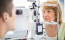
Retinal thinning appears to a consistent finding in patients with Parkinson‘s disease and questions have arisen over whether this serves as an early biomarker for the subsequent onset of the disease. Clinical writer Rod Tucker delves into the evidence.
Parkinson’s disease (PD) is estimated to affect at least 1% of people over the age of 60. The neurodegenerative condition affects both movement and cognition and is linked with the loss of dopaminergic neurons in the substantia nigra together with the presence of Lewy bodies.
Symptoms and progression of PD depend on the subtype, but patients with PD commonly experience tremor, bradykinesia and muscle stiffness, and up to 70% also have recurrent visual complaints and visual hallucinations.
But what is responsible for the visual dysfunction, and is it somehow linked to direct dopaminergic impairment within the eyes?
The fact that dopaminergic neurotransmission occurs within retina has been known since 1982, and the first evidence that patients with PD could experience visual problems emerged in 1987. Researchers observed a loss in both flicker sensitivity and near the peak of the spatial contrast sensitivity in those with the disease. But whether these visual deficits was causally related to PD remained unknown.
Definitive proof linking visual dysfunction with PD finally arrived, but from a rather unusual source. Canadian researchers decided to measure the level of dopamine in the retinas of eight patients with PD who died.
They stratified their findings based on each individual‘s last dose of levodopa therapy. While the retinal dopamine content in five patients who received levodopa therapy before death was similar to that in the controls, the levels were significantly lower in those who had not received the drug within the last five days before death.
As it became widely acknowledged that retinal levels of dopamine were often reduced in patients with PD, whether this was also associated with a concurrent loss of retinal tissue was less clear. Establishing such a relationship became much easier in 1994, following the introduction of optical coherence tomography (OCT).
This technique, which uses near-infrared light, enabled an assessment of nerve fibre thickness in the eye. In 2004, using OCT, researchers were able to show there was reduced thickness of the circumpapillary retinal nerve fibre layer in patients with PD.
Later work quantified this reduction in comparison to healthy controls and established that retinal thinning of the inner retinal layer was significantly greater in those with PD.
Although there was an emerging consensus that retinal thinning was a hallmark of PD, it wasn‘t actually a universal finding. For instance, other work was unable to detect a difference in comparison to healthy controls. A second study, which also failed to detect differences with PD and healthy controls, called for further work to define the role of OCT as a diagnostic biomarker.
Nevertheless, in a 2018 study, Korean researchers not only re-affirmed the presence of retinal thinning in patients with PD but provided the first evidence that there was a correlation between retinal thinning and nigral dopaminergic neuronal loss within the brains of patients who were at an early stage of their disease.
The relationship between retinal changes and PD was further strengthened in a 2021 meta-analysis of 27 studies that included 1,470 patients. The authors concluded that whole thickness – the thickness of the combination of ganglion cell layer and inner plexiform layer – and nerve fibre layer of central retina are significantly reduced in PD patients.
With confirmation of the relationship between retinal thinning and nigral dopaminergic neuronal loss, researchers sought to address a further pertinent question. If retinal thinning could be detectable in those without overt PD, could it serve as a prognostic marker for the disease?
This was the question posed in a recent study in the journal Neurology. Researchers assessed retinal thinning in two separate cohorts: one in patients with established PD and one in a second prospective cohort where retinal imaging had been performed.
The team looked at several aspects of retinal tissue including the ganglion cell-inner plexiform layer (GCIPL) and inner nuclear layer (INL) thicknesses, which had been measured in the two cohorts with OCT.
As expected, individuals in the first cohort with prevalent PD had thinner GCIPL and INL when compared to healthy controls. But what was more intriguing was how, in the prospective cohort in which patients had undergone OCT imaging approximately 2,653 days before the onset of PD, there was also significant GCIPL and INR thinning in those who went on to develop PD.
For example, the hazard ratio for a thinner GCIPL was 0.62 per standard deviation increase. In other words, for each standard deviation increase in GCIPL thickness, the risk of developing PD was reduced by 38%. In fact, the relationship remained significant even after excluding patients who developed PD within the first 24 months after their retinal imaging.
Taken together, the findings from the current study suggest that retinal thinning can be detected almost seven years before the onset of PD.
With the recognition that visual dysfunction in patients with PD is a harbinger of cognitive decline and greater white matter damage over time, it seems that the retina could become a potential imaging target.
There is still much more work that needs to be done, in particular, establishing the sensitivity and specificity of these changes, before the results can be used to predict whether an individual will develop PD. But if retinal thinning does turn out to be an effective biomarker, it could be used to support the early diagnosis of PD, thereby allowing for more effective treatment planning in those deemed to be at risk of developing the condition.
14th April 2023
Measuring serum endocan levels could be a useful way to determine the extent of ulcerative colitis in patients according to a study by Turkish researchers.
Ulcerative colitis (UC) is an inflammatory bowel disease in which there is mucosal inflammation. The British Society of Gastroenterology have provided guidance on the classification of the extent of disease. Where inflammation starts in the rectum and extends to involve the whole colon it is termed pancolitis. If the inflammation extends from the distal to the splenic flexure, it is known as left-distal UC. Endocan (EC) is a circulating proteoglycan, that has involvement in immunity, inflammation, and endothelial function. It could serve as a biomarker for inflammation but its potential for evaluation and monitoring in UC remains uncertain and was the aim of the current study.
The researchers recruited UC patients diagnosed clinically and endoscopically. In addition, none of those in the UC group were using any treatments. The team also included a control group, without systemic disease or use anti-inflammatory drugs. All patients had blood samples taken for measurement of CRP (C-reactive protein) and EC.
Endocan measurement and extent of ulcerative colitis
The study had 65 patients in total, 35 of whom had UC. There was a significant difference in CPR and EC levels between the UC and control patients (both p < 0.001). Significant differences were also present with endocan and CPR for the pancolitis and left-distal UC groups (p < 0.001). Furthermore, CPR and endocan levels showed a significant correlation (r2 = 0.54) in those with pancolitis. A cut-off value of endocan of 95.2 pg/ml could be used for the detection of diffuse colitis (p < 0.001).
Citation
Albayrak B et al. A novel inflammatory marker for extensive ulcerative colitis; Endocan. BMC Gastroenterol 2023
13th January 2023
A brain-derived tau biomarker which can be easily measured in blood samples, represents an important step in the specific identification of Alzheimer’s disease-type neuro-degeneration according to a study by an international research group.
Biomarkers for the identification of Alzheimer’s disease (AD) are based on the A/T/N” system where ‘A’ refers to the value of a β-amyloid biomarker, ‘T’ the value of a tau biomarker and ‘N’, biomarkers of neuro-degeneration or neuronal injury. To date, at least two of these markers are satisfactory. For example, plasma β-amyloid marker measurement (A), accurately determines amyloid positron emission tomography status in cognitively normal research participants.
In addition, blood-based tau markers (T) could be used as a simple, accessible, and scalable test for screening and diagnosis of Alzheimer’s disease. Nevertheless, neuro-degeneration (N) markers such as plasma neurofilament light chain (NfL), which measures axonal injury, showed no discriminatory power for AD compared to other neurological diseases.
Rather than focusing on developing a more specific ‘N’, researchers recognised that since tau measurements can be contaminated by peripherally generated (i.e., from liver, kidney or heart), it might be better to focus to identifying a specific brain-derived tau. In the present study, researchers created an anti-tau antibody which was designed to selectively bind with only brain-derived tau and which would hopefully be specific for patients with Alzheimer’s disease. The team then set out to validate the biomarker in five independent cohorts, including autopsy samples.
Brain-derived tau and Alzheimer’s disease
Using paired cerebrospinal fluid (CSF) and serum samples from AD patients and controls, there was a strong correlation (Spearman’s rho = 0.85, p < 0.0001) between the brain-derived (BD) tau levels in serum and CSF samples. In contrast, serum and total tau measurements were no significantly correlated (Spearman’s rho = 0.23, p = 0.3364). In addition, plasma brain-derived tau accurately distinguished autopsy-confirmed Alzheimer’s disease from other neurodegenerative diseases, with an area under the curve (AUC) value of 86.4%.
Furthermore, using samples from patients in two memory clinic cohorts, serum brain-derived tau differentiated Alzheimer’s disease from a range of other neurodegenerative disorders (AUC = 99.6%). In another cohort, BD-tau levels were significantly increased in AD patient serum and CSF samples compared to controls (p < 0.0001). Finally, BD-tau levels were inversely correlated with clinical dementia rating global scores (Spearman’s rho = -0.30, p = 0.0352).
Taken together, the authors wrote ‘across cohorts, plasma/serum brain-derived tau was associated with CSF and plasma AT(N) biomarkers and cognitive function. Brain-derived tau is a new blood-based biomarker that outperforms plasma total-tau and, unlike neurofilament light, shows specificity to Alzheimer’s disease-type neuro-degeneration.’
They concluded that the brain-derived tau demonstrates potential to complete the AT(N) scheme in blood, and will be useful to evaluate Alzheimer’s disease-dependent neurodegenerative processes for clinical and research purposes.
Citation
Gonzalez-Ortis F et al. Brain-derived tau: a novel blood-based biomarker for Alzheimer’s disease-type neurodegeneration. Brain 2022.
1st September 2022
The presence of early onset colorectal cancer can be diagnosed non-invasively using an assay based on a plasma micro RNA (miRNA) signature according to the results of a study by an international group of researchers.
The World Health Organisation estimates that in 2020, there were 1.93 million cases of cancer of the colon and rectum (colorectal cancer) and which led to 916,000 deaths. However, there is some evidence to suggest that the overall incidence of colorectal cancer (CRC) is reducing but appears to be increasing in those under 50 years of age.
In fact, approximately 1 in 10 new diagnoses of CRC, termed early onset CRC (EOCRC), are now made in individuals 50 years or younger. Furthermore, there is a suggestion that early-onset carcinomas commonly show pathologic features associated with aggressive behaviour.
Consequently, it is necessary to develop a means of testing for ERCRC since a delay in the diagnosis could have a significant impact, leading to an early death.
There is accumulating evidence that circulating micro-RNAs, which are a class of non-coding RNAs that play important roles in regulating gene expression, also reflect physiological and pathological alterations in cancer patients and may serve as important surrogate minimally-invasive biomarkers.
It has already been shown that the miRNA, MiR-92 is significantly elevated in the plasma of patients with CRC and could become a potential non-invasive molecular marker for CRC screening. However, to date, there are no studies examining the potential value of miRNAs in patients with early onset colorectal cancer and in the present study, researchers set out to try and identify a suitable miRNA signature for this group of patients.
Early onset colorectal cancer and miRNA signature
The team identified 4 miRNA signature in patients with EOCRC and which had a diagnostic area under the curve (AUC) of 0.82 with a sensitivity of 0.72 and specificity of 0.84. The team tested the diagnostic potential of this 4 marker panel in plasma samples in both a training and validation cohort.
The validation cohort included 142 patients, 77 of whom had a median age of 43 years (48.1% female) and 65 control patients. The AUC was 0.88 (95% CI 0.82 – 0.93, p < 0.01) and which provided a sensitivity of 0.82 and a specificity of 0.86 and an accuracy of 84%.
In addition, the AUC for the biomarker panel was 0.92 for EOCRC in stages I/II and 0.87 for stages III/IV and could distinguish between early and later stage disease.
In an effort to show that the use of the miRNA signature was in fact discriminatory, the researchers measured levels in plasma samples of patients before and 3 months after curative surgery and found that there was a significant decrease in expression in the post-surgical samples (p < 0.05).
Finally, the team showed that the miRNA signature could also discriminate between patients with late onset CRC from non-disease control patients with an AUC of 0.84, a sensitivity of 0.82 and a specificity of 0.88.
The authors concluded that this novel miRNA signature could be used to detect patients with early onset CRC and that with further validation, could be used to transform the screening of patients with EOCRC and subsequently reduce mortality rates.
Citation
Nakamura K et al. A Liquid Biopsy Signature for the Detection of Patients with Early-Onset Colorectal Cancer Gastroenterology 2022
6th January 2022
A potential biomarker which indicates the presence of major depression and its response to treatment could become a useful diagnostic tool and create a platform for personalised medicine. This was the conclusion of a proof-of-concept study by a team led by researchers from Signant Health, Boston, USA.
Depression is the commonest mental illness worldwide and the incidence has increased by 49.8% from 1990 to 2017, affecting some 25.8 million people. Moreover, the incidence of major depression also appears to be on the increase, with a US study in 2021 indicating that number of US adults with major depression aged 18-34 years increased from 34.6 to 47.5% between 2010 and 2018.
Although treatment of depression with antidepressant drugs has become widespread, some evidence from a real-world study suggests that only a third of patients achieve remission with the antidepressant, citalopram. A potential biomarker that not only identified those with major depression but also enabled tracking of a patient’s response to treatment could therefore be invaluable to clinicians.
Studies suggest that the cyclic adenosine monophosphate (cAMP) cascade is down regulated in major depression but up regulated by antidepressant treatment. Moreover, G protein coupled receptors and their attendant G proteins and effectors, such as adenylyl cyclase, are involved in the generation of cAMP.
Many neurotransmitter receptors, G proteins, and signalling effectors such as second-messenger-generating enzymes are present in lipid rafts and one particular protein, G-s alpha (GSA), appears to be the target of antidepressants, which facilitate the activation of adenylyl cyclase by making GSA more available.
In the present study, researchers hypothesised that antidepressant treatment increases the concentration of GSA, enabling it to exit from the lipid raft and stimulating the enzyme, adenylyl cyclase. They used blood platelets to determine adenylyl cyclase activity based on its response to prostaglandin E1 (PGE1) stimulation, which served as their potential biomarker by measuring of the extent of GSA-adenylyl cyclase coupling in both patients with major depression and healthy controls. The level of depression was assessed using the Hamilton rating scale for depression (HamD) and included patients were required to have a score > 15 at the initial visit.
Findings
Using samples from 41 individuals with major depression and 44 healthy controls, baseline measurement revealed a significantly lower PGE1 activation in those with depression compared to controls (p = 0.02), suggesting that there was less GSA available.
A total of 19 depressed individuals (6 men and 13 women) with a mean age of 50.6 years and a mean baseline HamD score of 20.4 consented to having a course of antidepressant therapy. After 6 weeks of treatment, there were 11 responders (HamD scores > 50% improvement from baseline) whose platelet samples demonstrated a significant increase in PGE1 stimulated adenylyl cyclase compared to non-responders (p = 0.05).
In fact, treatment responders showed a 62% increase in mean PGE1 stimulation compared to baseline, compared to -4.6% decrease for non-responders.
The authors concluded that the translocation of GSA from lipid rafts, as reflected by an increased PGE1 stimulated adenylyl cyclase, after treatment with antidepressants, could be used as a potential biomarker for both major depression and patients response to therapy and called for future studies to determine the utility of this biomarker.
Citation
Targum SD et al. A novel peripheral biomarker for depression and antidepressant response Mol Psychiatry (2022).
29th November 2021
The total bile acid concentration among those with visible coronary artery disease (CAD) as seen with coronary angiography was approximately 50% lower than in patients without CAD, indicating that this might serve as an important biomarker for the disease. This was the finding of a study by researchers from the department of Cardiology, Cochin Hospital, Paris, France.
Cardiovascular disease is the leading cause of death globally, accounting for an estimated 17.9 million deaths every year. Moreover, other evidence shows that in patients with non-alcoholic fatty liver disease (NAFLD), who have an excessive fat deposition in the liver, there is a dysregulation of bile acid (BA) metabolism. Finally, one systematic review in 2013, revealed how NAFLD is a strong independent predictor of cardiovascular disease. Taken together, these findings might suggest that dysfunctional BA metabolism could be indicative of CAD.
For the present study, the French team sought to explore the relationship between total bile acid concentration and CAD by measuring the level of acids in patients referred for coronary angiography and who could well have CAD. In addition, the team also measured the concentration of 7-alpha C4, which serves as a marker for the production of bile acids by the liver to determine whether there was any correlation with CAD.
Findings
A total of 80 patients with a mean age of 61.5 years (61% male) were recruited, 45 who presented with significant CAD on angiography and the remainder who were normal. Both groups were comparable apart from the CAD group being significantly older (66 vs 57 years, p = 0.004), including more men (73% vs 49%, p = 0.02) and with a significantly higher diastolic pressure (77 vs 72 mmHg, p = 0.04).
After adjustment for age and gender, the total bile acid concentration was highly predictive of CAD, with an area under the curve of 83.6% (95% CI 74.5% – 92.8%) and an odds ratio, OR of 0.51 (95% CI 0.31 – 0.85, p = 0.01). In other words, bile acid levels were approximately 50% lower in patients with CAD. From a total of 28 different bile acids, the researchers identified that one particular BA, GCDCA acid, was also predictive of CAD (AUC = 83.6%) and with a similar odds ratio (OR = 0.60, 95% CI 0.01 – 0.51, p = 0.01). A further observation was how the levels of 7-alpha C4 was similar in both groups (0.13 vs 0.12 micromol/L). One interesting finding was how among 17 patients who had been prescribed statins, the mean concentration of total bile acid, doubled (0.68 to 1.37 micromol/L, p = 0.01) after one month of treatment.
In discussing these results, the authors suggested that the total bile acid concentration might serve as a natural braking mechanism and which protected against the development of atheromatous plaques. They concluded by proposing that GCDCA could serve as a biomarker and predictor of visible atheroma in patients with CAD.
Citation
Nguyen CC et al. Circulating bile acids concentration is predictive of coronary artery disease in human Sci Rep 2021
8th June 2021
Urothelial carcinoma (UC) or transitional cell carcinoma, is the most common form of bladder cancer and is the tenth most common cancer in the world. The urothelial cells that line the bladder and urinary tract are constantly exposed to environmental agents that are filtered through the kidneys and not surprisingly, 90% of bladder cancers arise in these cells. Detection of UC is based on subjective microscopic features that are not always able to distinguish between benign and low-grade UC. Now a team from Yale Cancer Centre, US have developed a urine screening test based on immunocytochemistry and detects keratin 17, a molecule that has been found to be over-expressed in a range of cancers such as those affecting the breast, ovaries and pancreas. The researchers wanted to examine whether identification of keratin 17 could be used to screen for UC and detect recurrent UC in urine samples.
The study involved two patient cohorts: one with haematuria and one with recurrent UC. In screening samples with haematuria, the test was found to have a sensitivity of 100% and specificity of 83%. In patients with recurrent UC, the corresponding specificity and sensitivities were 92% and 100%.
The test itself, URO17 is developed by KDx diagnostics and commenting on the results of the study, co-author, Dr Nam Kim, said “there is now a growing body of evidence that the non-invasive K17 urine test will make a significant positive impact on detection and management of UC”. The study authors concluded that the study provides evidence to support the development of prospective trials to further define the clinical and diagnostic impact of K17.
Source
Babu S et al. Keratin 17 Is a Novel Cytologic Biomarker for Urothelial Carcinoma Diagnosis. Am J Clin Pathol 2021
IMPORTANT LINKS
MORE FROM COGORA
© Cogora 2025
Cogora Limited. 1 Giltspur Street, London EC1A 9DD
Registered in the United Kingdom. Reg. No. 2147432