This website is intended for healthcare professionals only.
Take a look at a selection of our recent media coverage:
8th July 2021
Professor Sidhu described how King’s is a large tertiary, general hospital in southeast London serving a population of over 2.2 million people in a largely deprived area. King’s has several different sites that cover various services, including paediatric, neonatal, cardiothoracic, neurosurgery, breast cancer screening, and liver transplantation. There is one centralised, very large, radiology department employing around 80 consultant radiologists and up to 700 staff. The department is also involved in the provision of training for radiologists and radiographer staff. Although radiologists are qualified doctors, their path towards becoming a radiologist is a long one that can take up to 12 years. In contrast, radiographers, who are the individuals responsible for taking images, still need to undergo degree level training although their role has greatly expanded in recent years. At King’s for example, radiographers manage all aspects of imaging for CT and MRI scans imaging in interventional radiology and some radiographers have become sonographers involved in the scanning and issuing of medical reports. All aspects require several years of training to become established within these roles.
Ultrasound is available and employed in many different specialities by experienced and competent individuals. Professor Sidhu explained how there remains strict controls on who can perform scans that involve exposure to radiation and particularly in nuclear medicine, with the administration of radioactive isotopes. Administrators are required to have a specific ARSAC licence. All hospitals that undertake examinations with radiation exposure will have a radiation protection officer. In contrast, while there are no specific controls on who can perform either an MRI and ultrasound scan, he highlighted how MRI equipment is prohibitively expensive and ultimately is best reserved for the radiology department. Nevertheless, according to Professor Sidhu, the position is rather different with respect to ultrasound. While in the past, ultrasound has been the responsibility of the radiology department, as Professor Sidhu explained, things are rapidly changing with much ultrasound, particularly point-of-care, becoming performed outside of radiology. For instance, chest physicians will use ultrasound to identify pleural effusions that need to be drained, and rheumatologists will make use of ultrasound to examine the small joints of the hands. The evolution of ultrasound in other specialties has been considerable and radiologists no longer have either the time or perhaps inclination to perform such scans. Despite these developments, as Professor Sidhu described, it is safety regulations, focused on the patient, that keep radiology departments together. However, while the use of ultrasound has now extended beyond the realms of the radiology department, he felt that the radiology community was not overly concerned about this direction of travel, especially given that in the UK, there is a shortage of radiologists, and this delegation has to some extent been welcomed. In fact, he now believes that no single speciality effectively “owns” ultrasound and in many radiology departments, the ultrasound scanning is performed by sonographers and the radiologists themselves have moved on to becoming more focused on the interpretation of specialised scans. As a passing thought, he felt that while radiologists were likely to be the most competent individuals to perform and interpret scans, if an ultrasound was being used as a point-of-care service for a specific indication, provided that an individual healthcare professional appropriately interpret a scan, he had no objection to these developments.
Professor Sidhu mentioned how he had been a member of the European Society of Radiologists (ESR) ultrasound subcommittee for several years during which time, it had produced a number of different position papers. He cited what has become a very successful position paper on infection control and prevention from 2017,1 highlighting the importance of good hygiene measures, especially with transducers and how these should be cleaned after each use. More recently in 2020, the group have published best practice recommendations and imaging use.2
This latest paper was an update of an earlier 2009 position paper on ultrasound.3 As Professor Sidhu clarified, the newer version provides a series of recommendations on appropriate standards for the use of ultrasound in radiology from the perspective of the ESR. For instance, the position paper, defines the essential requirements for equipment, practice aspects of use, infection control, requirements for training, certification and competence. He noted that while not all ultrasound operators would necessarily conform to the standards delineated in the position paper, it did define the professional standards which would be expected if the service were delivered by a radiologist. Professor Sidhu explained that an important aim of the position paper was to hopefully clarify for any non-radiologists, the anticipated standards which should be followed, in a sense, a standard operating procedure for undertaking ultrasound, which had the support of the ESR and therefore credibility. Professor Sidhu described how it was important that the position paper provided the necessary guidance because when using ultrasound, it is not the machine which does the job but the operator. If the operator is not competent, neither is the output from the machine! In short, it is vital that operators will need to practice, learn and develop all the time to perfect the technique to ensure that they get the best use from the device.
In terms of ensuring continued best practice, Professor Sidhu outlined documents produced by the ESR were designed to support non-radiologist operators. This advice encouraged participation in audits of their service but additionally and equally important, was that non-specialists needed to maintain their skills and competence through an examination of the practice all the time, together will ensuring equipment maintenance, the environment used to perform imaging and how best to manage patient throughput, all of which were essential for adherence to professional standards. Professor Sidhu hinted that with a rapid pace of development in ultrasound technology, it is highly likely that the professional standards of today would require updating in the near future. He pointed to the fact that equipment was becoming smaller, so that handheld devices are able to do a reasonable job and ultrasound technology is also available for use on smartphones and an iPad for scanning.
Professor Sidhu indicated that one of his aims for the ESR over the next few years was to define the place of ultrasound within radiology more clearly. He remarked on how the position of ultrasound within radiology is far less clear cut than say 20 years ago and that is often seen as the ‘Cinderella’ modality in radiology. He noted for instance how today, a lot of younger radiologists were more interested in CT and MRI scanning because this was perceived as being more cutting edge and that ultrasound is often seen as harder work than the other modalities. After all, clinicians must operate the device, sit with the patient, and scan them, whereas there is no direct patient contact with MRI and CT, with the hard work coming from the ability to form a diagnostic image, readily interpretable. He revealed how for the next ESR meeting in 2022,4 the committee is preparing a session dealing with where they believe ultrasound will sit in 20 years’ time. He stated that there are plans for three speakers at the meeting and who will discuss different models of ultrasound practice. Firstly, there is the German model, in which many GPs perform ultrasound as well as having a centralised hospital department run by radiologists, but with other medical specialists effectively dipping into the service. Second is the Russian model, whereby both radiologists and other physicians only do ultrasound and no other imaging modality. The third, and often perceived as a controversial model, is the one deployed in the UK, where it is the radiologists who performs less scanning, taking on more specialised examinations (e.g. MSK) and delegating the sonographers to effectively undertake most of the scanning, and providing diagnostic reports. He felt that a discussion of the different models would undoubtedly provoke a lively debate. Professor Sidhu mentioned that tied in with this debate will be a position paper on where the ESR believes ultrasound should sit within radiology and how the speciality should evolve over the coming years. Professor Sidhu, though not the final arbiter on any decisions, felt that in the future, perhaps radiologists should not see themselves as guardians of the ultrasound world. He added that anyone from whatever subspecialty and who has an interest and can demonstrate competency and safety in their practice should be encouraged to use ultrasound. Professor Sidhu believed that there was nothing inherently wrong with, for example, a rheumatologist upskilled in the use of ultrasound, if they saw the benefit of the imaging modality in their assessment of a patient. He also thought it possible that radiologists could retain ultrasound within their departments but allowing access to physicians from different specialities with an overarching goal of improvements in patient care.
A further advantage of ultrasound highlighted by Professor Sidhu was the flexibility of the modality. In contrast to the static imagery of an MRI or CT scan, ultrasound was performed in real-time. Consequently, it was possible during the scan to enquire as to whether the patient experienced any pain or discomfort, particularly if the imaging indicated a potential cause for the pain. Alternatively, the patient may offer a snippet of history during the scanning, and which helps to confirm the diagnosis, neither of which are available to radiologists when interpreting other imaging modalities.
Professor Sidhu described how King’s was very busy during the first and second waves of the COVID-19 pandemic.5 With the global cancellation of routine imaging during the first wave, the radiology department was quiet with ultrasound becoming extremely useful within the intensive care unit as a point-of-care for imaging of patients’ lungs; this was done predominately by the pulmonary physicians although radiologists did perform some abdominal scans. He added that during the second wave, the department was a lot more prepared and continued with as much routine scanning, if patients could safely attend the hospital, but suspects that this has resulted in a huge backlog of imaging awaiting to be undertaken. While the magnitude of this backlog remains uncertain, Professor Sidhu remarked that there are still a lot of patients waiting to be scanned.
Professor Sidhu thinks that an important learning from the pandemic is that services need to become more patient centric and that the provision of imaging services should become easier and more accessible for the patient. With this idea in mind, there is likely to be a wholesale shift of routine outpatient scanning out of the acute hospital and make greater use of community-based imaging, a move he says which has been supported by government. He mentioned that although this change had been under discussion for several years, it was really brought into sharper focus as a consequence of the pandemic. After all, he reflected on how it has been ludicrous to bring large numbers of patients into a busy hospital for routine/GP imaging. Consequently, there is now a move in progress to establish diagnostic hubs, adjacent or close to the hospital and introducing agreed patient pathways to ensure that only those patients who need further management must visit the hospital. Professor Sidhu felt over the next few years, elective imaging could be undertaken within the hub and that this would release capacity with the hospital, allowing time to see acute patients and those who required other forms of interventional or complex imaging, with the important caveat, that the hospital service is accessible for patients when needed.
Professor Sidhu noted how there were enormous developments in ultrasound technology, and which were of huge benefit. He mentioned that a difficulty was that many people still see ultrasound as only black and white, but that this is no longer the case and ultrasound has a far greater capability for imaging that all other modalities. He cited how multiparametric ultrasound imaging can be used to assess patients with steatotic livers6 and that other innovations such as colour Doppler ultrasound, contrast ultrasound and elastography ultrasound,7 which looks at the stiffness of the liver, the amount of fibrosis and scarring have all proved to be of value in patient care. He added that a further advantage of the developments in ultrasound was that the technology was less expensive than other modalities and safe. He sensed that in the next couple of years there would be many further innovations with ultrasound-based technology and that these would continue to be patient-friendly. Using the example of scanning the liver of a two- or three-year-old child, Professor Sidhu described how for an MRI scan, the child needed to be sedated perhaps, given the contrast agent gadolinium, and kept still. However, for an ultrasound scan, the child remains awake, and the parents can also be present to help with any possible anxiety. He thought that as an imaging modality, radiologists would be ultimately unwise to give up on ultrasound, adding that the technology is now at a level where the device does almost everything for the operator, adjusting the parameters automatically to produce the best image.
Another development that had made a considerable impact on ultrasound mentioned by Professor Sidhu was artificial intelligence. The technology can recognise the organs under investigation, identifies any abnormalities as well as providing a differential diagnosis. He added that it even writes the report for the operator although currently, it still needs a clinician to interpret the results of the scan.
Professor Sidhu thinks that will all the emerging technologies, some radiologists have been left behind simply because the developments in ultrasound are largely driven by physicians in other specialties; in particular, hepatologists. He explained how a key driver is not so much that other specialists embrace the technology but more that unlike radiologists, hepatologists and other specialties do not have routine access to CT and MRI, ensuing the best aspects of ultrasound are utilised constantly, before reverting to another imaging modality.
Even though in the future ultrasound might well move outside of the sphere of radiology, Professor Sidhu still believes that there will always be a need for radiologists to be skilful in ultrasound. As a profession, they possess the necessary skills to match up the results from all the other scans and images and in doing, so will continue to make an important contribution to patient care.
There can be no one truly unaffected by the COVID-19 pandemic. This ongoing international health crisis has impacted on lives around the world; arguably, none more so than those of front-line health care workers. Speaking as a sonographer based in a large teaching hospital in the north of England, I have experienced first-hand the significant impact the pandemic has had on service delivery, my colleagues and, most of all, our patients. However, as an optimist, I like to believe that with every dark cloud a silver lining can be sought. Therefore, as an optimist, one opportunity the pandemic presents is the opportunity to reset normal within our clinical and professional landscapes. In some instances, of course, this happened rapidly and without time to pause and reflect. However, there has been an opportunity to review those services that were stopped and an opportunity to redesign as we move to restart planned care. Indeed, in my own institution, entire pathways have been remodelled, placing ultrasound imaging at the very core of where it was once an afterthought and poorly resourced or acknowledged. This has provided an ideal opportunity for modern ultrasound imaging to be showcased; those technological developments that have gradually become integral in diagnostic ultrasound imaging are now being recognised as essential to providing a powerful first-line core test for many patients, particularly those with delayed presentation and for whom swift assessments and management plans are required.
The British Medical Ultrasound Society (BMUS) has the core objectives: “to promote the advancement of the science and technology of ultrasonics as applied to medicine” and “to ensure the highest standards in practice are maintained”. Coupled with the changing role of ultrasound in patient pathways, BMUS has developed and provided education and guidance to all professionals working in the field of medical ultrasound. For BMUS, as the premier ultrasound society in the UK, the annual scientific meeting has been the stage to present developing best practice, new and emerging technology, and provide an opportunity to discuss research and development with like-minded professionals. Face to face events have clearly been impossible to hold during the pandemic, and difficult to consider planning in the immediate future. This, too, has led to the organisation pausing, reflecting, and redesigning what can be delivered. The need for support for ultrasound professionals has not diminished; indeed, with increasing role development, it can be argued the need is greater than before. A virtual webinar programme was developed by BMUS during 2020 to provide relevant, and pertinent, education and standard setting in line with the objectives of the organisation; a journal club via Twitter established, and now plans are well underway for a full virtual online conference. None of these are likely to have developed without the dark cloud of the COVID-19 pandemic causing much disruption.
Webinars and e-learning, in the world of medical ultrasound at least, have developed at a pace in the last 18 months. Having a creative and innovative team supporting an online training programme has been essential and has unearthed hidden talents within the BMUS administrative team as well as the multitude of professionals who have supported and volunteered to provide the education programme. As with many professional charitable organisations, BMUS is reliant on volunteers who are willing and able to share their time and knowledge with peers. Despite the difficulties we have all faced, the willingness of our clinical colleagues to support the ongoing education and professional guidance output of BMUS has been phenomenal, and yet, despite this wealth of readily available material, attracting new members and minimising attrition of existing members remains as very real challenge. It is pleasing to note that the sonographer membership of BMUS has marginally increased recently, but attracting medical colleagues, in particular radiologists, to support the organisation remains difficult. BMUS prides itself on being a multi-disciplinary organisation and there to support any professional using medical ultrasound within their scope of practice. In the UK, by far the largest professional group is sonographers who primarily use ultrasound in their everyday clinical practice. Essentially, a sonographer is medical ultrasound! Radiologists learn ultrasound as a core skill during their training but, and as is common in larger establishments, radiologists are specialty-focused and many use cross-sectional imaging modalities or interventional suites that are now far removed from the hands-on image acquisition, interpretation and reporting of ultrasound imaging. Increasingly, there are numerous non-radiology professions using medical ultrasound as a tool to enhance their clinical practice and to guide patient management. Indeed, many Royal Colleges now have specific ultrasound training programmes and competency assessments that their members are expected to complete to be able to deliver this point-of-care ultrasound (PoCUS). The importance of PoCUS cannot be overstated. As such, publications produced by our emergency medicine, chest, and intensive care colleagues ensured shared learning of the signs of COVID-19 on lung imaging, which became critical as hospital admissions rose in the first and second waves. The aim of BMUS is to bring this multi-disciplinary family together and recognise the benefits of sharing knowledge and skills but, at the same time, not alienating those for whom medical ultrasound is a professional career choice; sonographers, physicists, and radiologists alike.
BMUS has had, as do many societies, sub-committees whose roles are to ensure the objectives of the organisation are met. Longstanding and hardworking committees such as the physics and safety, publications and education committees have been the workhorses of the society since its inception. Latterly, new committees have been set up to reflect the changing professional landscape and, the now more diverse, users of medical ultrasound. PoCUS and obstetric clinical imaging groups have been established to support improvements in these critical fields of medical ultrasound. The professional standards group was established to better nurture and enhance working relationships with like-minded professional bodies and organisations. A consultant sonographer interest group has been established to promote career development for non-medical ultrasound practitioners and show case the very best in ultrasound innovation. However, membership of the society and an engaged workforce, from whatever background, is essential to the future success of these committees and the wider BMUS family. BMUS upholds the highest standards of practice and, it is with this aim, that the society engaged with Health Education England (HEE) in a project to increase the sonography workforce. BMUS, in collaboration with the Consortium for Accreditation of Sonographic Education, the Royal College of Radiologists and the Society of Radiographers has been integral member of this HEE sonographer workforce project group and progress is slowly being made. Every arm of health care is under pressure to increase activity, improve turn-around-times, and deliver safe and effective care. There is no profession that is not struggling with workforce capacity, with vacancy rates well documented; radiology and sonography are no different, and solutions have needed. While BMUS has no remit to act as a trade union or provide workplace advice, it does have a remit to ensure standards of practice are optimised, safe and of high standards. The multi-disciplinary nature of its membership ensures BMUS is well-placed to offer opinion, advice and guidance as part of this collaborative approach to HEE as the path of defining and developing an ultrasound / sonography career framework emerges.
Whilst there have been significant challenges, in the workplace, professionally and personally over the last 18 months there has been opportunity to reflect and set a new or revised course for the future. The publication of the Richards Report in 20201 and NHS Long Term Plan in 20192 has provided an opportunity for the role of sonography, and the role of medical ultrasound within healthcare, to be established as essential in first line investigations in critical, acute and planned care, the improvement of obstetric outcomes, and in guiding interventions within an out-patient setting. Professionals working with medical ultrasound, as a career path or using it as a tool, have the opportunity to embrace new ways of working and new pathways. Supporting national organisations, such as the BMUS, provides the opportunity for professional societies to have a voice around the table at national discussions and highlight the benefits of a highly trained, well-motivated, recognised workforce. There are challenges ahead but none that cannot be faced together.
I am proud to represent BMUS and my multi-disciplinary ultrasound colleagues and will work hard in my tenure as President to turn those challenges into golden opportunities as we head into the new normal of 2021 and beyond.
Pamela Parker is a consultant sonographer working within radiology of the Hull University Teaching Hospitals NHS Trust. Pamela has over 25 years’ experience in the field of medical ultrasound. Her specialist interests are uro-genital ultrasound, contrast and fusion-guided imaging. She has established the first sonographer-delivered fusion and transperineal prostate biopsy service within radiology in the UK. Pamela is studying part-time for a PhD investigating the role of ultrasound within the active surveillance of prostate cancer.
Dr Dugar’s department has approximately 16 radiologists and, with a newly purchased third CT scanner, the department is extremely busy, operating on an almost conveyor belt-like basis. In a typical day, radiographers will perform around 1000 imaging investigations including those for elective, acute and ward patients. As there is a requirement to have A&E CT scans reported on within an hour, Dr Dugar emphasised the importance of prioritising the workload within the team. Hence, one radiologist is always allocated to report emergency scans, and while others do elective work. During the evening and weekends only one radiologist is available for undertaking emergency CT and MRI scan reporting. Night-time emergency radiology has now been outsourced to Australia. Since starting her role over 17 years ago, the workload had expanded considerably due to a combination of increased expectations and national guidelines that often recommend the use of imaging. For example, she described how 17 years ago, during a typical Sunday, she might be called upon to report one emergency scan but on a recent weekend shift, her hospital performed 100 emergency CT and MRI scans. She feels that no other specialty has experienced that kind of explosion in workload.
Dr Dugar emphasises that technology is the backbone of radiology and had a desire to make best use of technological advances as a means of enhancing patient care. She explained that she was appointed as informatics advisor in 2015, because of an interest in the topic coupled with the fact that she had led the development of digitisation in her own department. She suggested that the Royal College of Radiologists (RCR) felt that it was necessary to have a committee that was able to set the standards of what should be achieved by all radiology departments implementing informatics. In other words, the RCR Informatics Committee was tasked with defining the standards and hence, best practice, which should be achievable through hospitals’ information technology (IT) systems.
Out of this work came the publication Integrating artificial intelligence (AI) with the radiology reporting workflows (see later in the supplement for a summary of guidance). The guidance defined the standards for how AI should be incorporated into the radiology information (RIS) and picture archiving and communication systems (PACS). Dr Dugar highlighted how, in some respects, AI is considered a very broad term and can be interpreted differently depending on the context. For the present document, the RCR informatics group considered AI in the narrow context of ‘computer vision’ used for radiology image pre-analysis. As Dr Dugar explained, because AI systems have already been developed for facial recognition, given that the role of a radiologist is to visualise images and to make interpretations to inform the ongoing care of patients, it seemed only right that this should be the area to focus upon.
However, a primary focus was to ensure that IT vendors could develop the necessary infrastructure to incorporate AI systems with different hospitals. A further consideration for the implementation of AI was the apparent national shortage of radiologists in the UK. For instance, in a 2018 report,1 it was noted how in the UK, only one in five UK Trusts and health boards had enough interventional radiologists to provide safe 24/7 services to perform urgent procedures.
Dr Dugar defined how the AI workflow guidance should work in practice, explaining that the AI system would initially review the image before it was seen by a radiologist. The AI system algorithms were such that it was able to highlight any relevant features, which is also within the remit of radiologist. Nevertheless, whereas the AI systems are capable of detecting some abnormalities, the radiologist would then combine these findings with other test/imaging results, and any other relevant clinical findings, to create a more personalised report for the patient.
If the AI system can do the essential job of a radiologist, surely these individuals can be easily replaced? Dr Dugar disagrees. She revealed how in 2016 AI pioneer, Geoffrey Hinton, had said “we should stop training radiologists now. It’s just completely obvious that within five years, deep learning is going to do better than radiologists.”2 At the time, she said this created a major staffing crisis, especially in the US, which saw a downturn in doctors choosing radiology as a career, fearing that they would be deemed superfluous in the near future. But, as Dr Dugar added, medicine is not maths – if it were then there would be no need for a radiologist! She feels that it is virtually impossible to create an ‘artificial’ radiologist simply because of the huge number of algorithms that would be required to emulate the thought-processing of a radiologist.
Dr Dugar believes that having an AI system evaluating a scan goes some way towards the need for two independent reviewers of a scan result. This is considered as best practice, lending support to the metaphor that “two heads are always better than one”. As she said, this is the current recommendation for breast cancer screening. She stressed that having two independent reviews was necessary because even though individual radiologists are highly trained, they are fallible. Thus, from a safety perspective, dual review is the ideal standard.
The integration of an AI system was also important but for a very different reason. Dr Dugar highlighted that, in reality and with a national shortage of radiologists, it becomes impossible to achieve the two-reporter standard for images. One of the key reasons for a second reporter was to the minimise the phenomenon of satisfaction of search, which describes the situation where some lesions remain undetected after an initial lesion. As Dr Dugar illustrated, when a radiologist finds an abnormality, they become fixated on that particular problem and start to process this finding within the context of other clinical information and sometimes ignore other findings. With an AI system able to review the image prior to the radiologist, it effectively becomes that second reporter and a helper, alerting the radiologists to the full range of abnormalities present on the image. In discussion with colleagues, a barrier to greater use of AI is the perception among some radiologists that the system is very sensitive but not specific. Using the example of the assessment of a lung scan, Dr Dugar explained that while the AI system would report on the presence of tiny nodules, the focus of the radiologist was in looking for metastases. With greater knowledge of the patient’s clinical history than the AI system, the radiologist can potentially discount the relevance of these nodules and advise accordingly. In contrast, the AI system simply identifies any abnormality and is unable to make a subjective judgement within the context of any other clinical findings. Dr Dugar labelled the AI system as a ‘junior radiologist’, i.e., it was able to provide a limited role as a preliminary reviewer on images. Nonetheless, she did believe that in the future, with improvements in AI occurring, the input from a radiologist might be unnecessary, especially for simple imaging, e.g., reporting on the presence/absence of a fracture. However, radiologists would still be required to interpret more complex imaging from MRI or CT scans. She added that while an AI system could identify a filling defect in the lungs and report the most likely cause to be a pulmonary embolism, as a radiologist, you are always thinking laterally about other differential diagnoses.
As Dr Dugar described, being both medically qualified and trained in radiology allowed her and her colleagues to assess whether or not a particular imaging request was appropriate. She emphasised how often both junior doctors and those from other specialities, may not be completely clear on which imaging tests were correct. The vetting (triaging) and cancellation of inappropriate radiology requests document was introduced simply to help manage the workload within radiology departments. An important part of Dr Dugar’s role is to always vet or triage any requests for imaging that reach the department. This vetting process, she added, was crucial because of the high workload of the department, which makes it impossible to perform every exam request.
While the vetting process amounts to a clinical assessment task in itself, Dr Dugar highlighted how within her department over 90% of the vetting process was undertaken by the radiographers rather than the radiologists. This had been made possible through the introduction of a protocolised approach for the radiographers. Moreover, Dr Dugar believes that radiographers can quickly acquire the necessary vetting skills and then approve and book a scan or reject the request. A further advantage to radiographer-based vetting, is that, as these individuals are involved in performing the imaging, they are able to quickly assess, and then cancel, any duplicate requests and in some cases, even determine if the request would be of additional value, i.e., if the request is for a broadly similar scan. A difficulty for radiographers, however, is that without the necessary medical training, they may feel uncomfortable cancelling an imaging request that was requested on clinical grounds and in such instances, the protocol would dictate that the request is forwarded to the radiologist. As a consequence of introducing the vetting process in her department, Dr Dugar felt that on a typical day, she might be asked to vet up to 30 requests and allocates up to 30 minutes of her day to this task. She thinks that such vetting is a key task given that the department performs around 1000 scans each day. Although some of the requests passed to her from radiographers can be challenging, in many cases, it can sometimes be very straightforward and require simply altering the request to a more appropriate imaging modality. For more complex cases, she will need to review the patient’s medical history or initiate a discussion with the referring clinician to discuss the best option. In cases where the request is rejected, Dr Dugar ensures that the requesting clinician is informed of her decision and the rationale behind the cancellation. She thinks that the departmental system, being fully electronic has streamlined the whole request/cancellation process.
Dr Dugar expressed the view that the vetting document was desperately needed because in some radiology departments, no vetting process was in place. She realised that part of the reason behind this lack of vetting was largely due to a lack of functionality within the hospital’s internal IT system. The purpose of the vetting guidance was thus to ensure that while not all NHS Trusts employed the same vendors, these vendors would modify the IT infrastructure to enable electronic communication for the vetting process. As Dr Dugar said, in discussion with radiologists from outside of her own department, the cancellation process was often not communicated to the original requesting clinician and this led to internal friction and, in some cases, the radiologists in an attempt to appease the requesting clinicians, decided to no longer triage requests, with a resultant increase in their workload. Although it seems unusual that the whole of the NHS must deal with different IT vendors, Dr Dugar is against the idea of a national vendor. She thinks that with such a huge monopoly, there would be little incentive to innovate. What is more important she feels, is that the same workflow processes should be adopted in the different NHS Trusts, to improve the efficiency of radiology departments, even if this occurs through dissimilar IT systems.
Dr Dugar said that for her, radiology services did not stop during the pandemic although the focus shifted to COVID patients. She felt that her own work, which is either cancer or emergency-based, did not slow during the pandemic. She believed that one of the greatest changes because of the pandemic was the digital transformation within the NHS. She thinks this was of enormous benefit, enabling more virtual meetings which were a great advance compared to teleconferences. Another important development for work–life balance was allowing radiologists to have workstations at home. Dr Dugar says that having an interest in digital technology, she had tried to implement greater home working for some time, but her request was always denied due to lack of funding. However, she also thinks in the future, this innovation of homeworking and virtual meetings will not revert to pre-pandemic times but there will still be a balanced need for office working.
Dr Dugar believes that AI algorithms will develop in the next two to five years and become a much better preliminary reporter on many more things such as fracture detection, lung nodule detection etc. She mentioned how AI is already being used in brain imaging for strokes. She worries, however, that future innovations in AI by computer scientists will require additional funding, and that this should not be at the expense of a radiologist training.
Another potential growth area she feels is in the evolution of enterprise imaging3 and revealed how all her own radiology department’s images have already been archived and made available throughout the enterprise. An important current problem, she explained, was how various medical images from other specialties/departments have been generated but are stored in different locations and formats without the correct patient identifiers etc. and are not indexed properly (e.g., endoscopy images, ECG, audiometry, sleep studies etc). Incorporation of all the images and graphs in a single and accessible location will be of enormous value, not only to radiologists but also all treating physicians. With the ability to review all images, and together with other pieces of clinical data, it will allow radiologists to create a much more personalised report for the patient.
Although improvements in smartphone technology allow for image review, Dr Dugar thinks that at the present time, the quality of the images is not of sufficient quality for diagnostic purposes. Furthermore, from a medico-legal perspective, she would not use the images reviewed on a smartphone for reporting. She also felt that while mobile scanning units for MRI and CT scans were available and could be utilised for elective work, these imaging modalities would still need to remain within the hospital premises, where the equipment was needed for emergency scans.
Image acquisition and interpretation were separate, and Dr Dugar believes that she does not need to be present at a mobile scanning unit and can remain either in the office or at home to undertake her interpretive role for the images. However, radiologists (whether remote or on-site) must continue to work closely with the radiographers operating the scanners to provide support and advise on appropriateness and vetting/triaging support. Radiologists and radiographers must always work as teams to improve patient care.
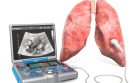
While computer tomography (CT) has become the mainstay of diagnostic imaging evaluation of thoracic disorders, in the hands of an experienced radiologist, lung ultrasonography has much to offer according to guidance issued by the European Radiology Society (ERS).
Within an intensive care setting, for example, point-of-care ultrasonography enables direct, bedside examination of the lung and pleural space and has been found to reduce the need for chest radiographs and CT scans. Although the standard of care within an intensive care unit is the chest X-ray, in an assessment of the comparative diagnostic performance of auscultation, chest radiography and lung ultrasonography, the latter was deemed to be highly sensitive, specific and reproducible for diagnosing the main lung pathologic entities in patients with acute respiratory distress syndrome (ARDS), which is a recognised complication of infection with COVID-19. More recently, compared with X-rays and CT scans, point-of-care ultrasound of the lungs of patients with COVID-19 has been found to offer similar performance to CT and superior to X-ray, in evaluating pneumonia and ARDS. In fact, the authors of this latter study commented on how lung ultrasound played an important role within the emergency room and intensive care unit, reducing exposure to radiation and minimised transport of high-risk patients.
With the potential benefits of ultrasonography clearly defined, the ERS has released, what it hopes, will be the definitive guide on the role of lung ultrasound in patients with COVID-19. The guidance covers the fundamentals of ultrasonography examinations, the different types of available transducers, including appropriate methods of cleaning and system set-up and concludes with advice on how to perform a lung examination and examples of the typical findings seen in patients with COVID-19.
The guidance defines how when using ultrasound for the diagnosis of lung pathologies, the presence of two artefacts can provide invaluable information. In fact, the document provides several illustrative imaging examples. The first, described as “A-lines” are reverberation artefacts triggered by oscillating tissue with an air interface and which can be seen as parallel, repetitive horizontal lines of the pleural surface. This A-profile is shaped by intact (‘dry’) lung parenchyma containing air when it is combined with normal lung sliding. If sliding is absent, however, it is intensely suggestive of a pneumothorax. In contrast, “B-lines” are vertical hyperechoic artefacts and arise from areas of pleural consolidation and are indicative of accumulation of fluid in the pulmonary interstitial space or alveoli. According to the ERS guideline, although one or B lines might be considered as normal, any increase in number or spread in one zone represents severe pulmonary interstitial oedema.
In a discussion of the different transducer devices, e.g., high and low frequency linear, curvilinear devices, the guidance suggests that high-frequency probes will offer a better solution to observe lesions in the pleural line. Nevertheless, it also makes the important point that ultimately, the performance and interpretation of results is not probe-specific. With respect to cleaning, the guidance indicates that though immersion of the probes in a strong disinfectant is desirable, in the longer term, repeated cleaning could damage the probe.
In fact, the recommendation is that the use of advanced cleaning solutions should be discussed with local infection teams and vendors supplying the machine.
The document ends with some general advice on basic lung examination and the typical findings observed in patients with COVID-lung disease. It notes how pleural effusions are uncommon and that these are more likely among those who are critically ill. The guidance records how lung ultrasonography can reveal the typical patterns for interstitial pneumonia, which in COVID-19 is mainly seen in the peripheral pulmonary zones.
Finally, the guidance notes that while CT may be needed for follow-up of cases in which ultrasonography is unable to provide a specific answer, the portability of the imaging modality avoids the need to transport high-risk patients while at the same time, can provide a prompt and accurate evaluation of the severity of COVID-19 pneumonia as well as permitting tracking of the disease during follow-up.
Citation
European Society of Radiology. The role of lung ultrasound in COVID-19 disease. Insights Imaging 2021. https://doi.org/10.1186/s13244-021-01013-6
Download Hospital Healthcare Europe free app
© Cogora 2024
Cogora Limited. 1 Giltspur Street, London EC1A 9DD
Registered in the United Kingdom. Reg. No. 2147432