This website is intended for healthcare professionals only.
Take a look at a selection of our recent media coverage:
14th August 2023
Use of machine learning has enabled scientists to accurately predict four subtypes of Parkinson’s disease based on images of patient-derived stem cells.
Parkinson’s disease is a neurodegenerative condition that affects both movement and cognition. Symptoms and progression vary based on the underlying disease subtype, although it has not been possible to accurately differentiate between these subtypes.
This may well change in the near future as a team based at the Francis Crick Institute and UCL Queen Square Institute of Neurology, together with the technology company Faculty AI, have shown that machine learning can accurately predict subtypes of Parkinson’s disease using images of patient-derived stem cells.
The work, which was published in the journal Nature Machine intelligence, generated a machine learning-based model that could simultaneously predict the presence of Parkinson’s disease as well as its primary mechanistic subtype in human neurons.
In the study, researchers generated stem cells from a patients’ own cells and chemically created four different subtypes of Parkinson’s disease: two involving pathways leading to toxic build-up of the protein α-synuclein and two involving pathways leading to defunct mitochondria. Together, this created a ’human’ model of the disease.
Next, researchers imaged these disease models and ‘trained’ the machine learning algorithm to recognise each subtype, from which it was then able to predict the particular subtype.
The machine learning model enabled researchers to accurately identify a disease state from a healthy control state.
With quantitative cellular profile-based classifiers, the models were able to achieve an accuracy of 82%. In contrast, image-based deep neural networks could predict control and four distinct disease subtypes with an accuracy of 95%.
The machine learning-trained classifiers were able to achieve a level of accuracy across all subtypes, using the organellar features of the mitochondria, with the additional contribution of the lysosomes, confirming the biological importance of these pathways in Parkinson’s disease.
James Evans, a PhD student at the Francis Crick Institute and UCL, and co-first author with Karishma D’Sa and Gurvir Virdi, said: ‘Now that we use more advanced image techniques, we generate vast quantities of data, much of which is discarded when we manually select a few features of interest.
‘Using AI in this study enabled us to evaluate a larger number of cell features, and assess the importance of these features in discerning disease subtype. Using deep learning, we were able to extract much more information from our images than with conventional image analysis. We now hope to expand this approach to understand how these cellular mechanisms contribute to other subtypes of Parkinson’s.‘
Sonia Gandhi, assistant research director and group leader of the Neurodegeneration Biology Laboratory at the Francis Crick Institute, who was also involved in the study, said: ‘We don’t currently have treatments which make a huge difference in the progression of Parkinson’s disease. Using a model of the patient’s own neurons, and combining this with large numbers of images, we generated an algorithm to classify certain subtypes – a powerful approach that could open the door to identifying disease subtypes in life.
‘Taking this one step further, our platform would allow us to first test drugs in stem cell models, and predict whether a patient’s brain cells would be likely to respond to a drug, before enrolling into clinical trials. The hope is that one day this could lead to fundamental changes in how we deliver personalised medicine.‘
Criteria to define both complete and partial clinical remission in patients with severe asthma has been developed by the Severe Asthma Network Italy (SANI) group.
Published in the Journal of Allergy and Clinical Immunology in Practice, the SANI group set out to highlight a consensus for asthma remission using the Delphi technique.
A panel of 80 experts, which included pneumologists and allergists from the SANI network, covering 57 severe asthma centres, and more then 2,200 patients, were included in the Delphi process.
In the first Delphi round, the group created 32 statements, which were divided in four main categories: general questions about remission; criteria for clinical remission criteria; complete or partial clinical remission and its duration; and cut-off values of different scores regarding disease control, lung function and inflammation. Each of these statements used a five-point Likert scale to measure panellist’s agreement to each statement.
The statements were sent to the panellists and, following an interim analysis, the responses were discussed to produce a consistent questionnaire for the second round.
During the second Delphi round, the criteria for complete clinical remission was confirmed as a composite of the absence for the need for oral corticosteroids (OCS); the absence of symptoms, exacerbations or attacks; and pulmonary stability. Moreover, these criteria had to be present for at least 12 months.
In contrast, partial clinical remission was defined where there was no further need for using
OCS, plus two out of the following three criteria: absence of asthma symptoms, absence of asthma
exacerbations or attacks, and pulmonary stability.
‘This SANI Delphi analysis defined a valuable, independent and easy-to-use tool to test the efficacy of different treatments in patients with severe asthma enrolled into the SANI registry,‘ the authors said.
Approximately 10-20% of people with asthma are estimated to have severe disease. The SANI group hopes that its definitions of complete and partial remission can be used to test the efficacy of different
treatments in patients enrolled and followed in the SANI registry.
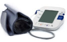
Using the incorrect cuff size when taking blood pressure measurements can lead to significant over and under-estimations, according to the findings of a randomised trial by US researchers.
Although clinical practice guidelines recommend the selection of an appropriately sized cuff based on mid-arm circumference before taking a blood pressure (BP) reading, the extent to which miscuffing affects readings was previously uncertain.
Now, in a study published in the journal JAMA Internal Medicine, researchers from Johns Hopkins University School of Medicine in Baltimore, Maryland, revealed that miscuffing results in ‘strikingly inaccurate‘ BP measurements.
In the randomised crossover trial of community-dwelling adults with a wide range of mid-arm circumferences, the researchers determined the effect of using a regular BP cuff versus an appropriately sized BP cuff on automated BP readings. Participants underwent four sets of triplicate BP measurements, with the initial three sets using an appropriate, too-small or too-large BP cuff in random order. In the fourth and final set of triplicate measurements, an appropriate BP cuff was used.
The team set the primary outcome as the difference in mean blood pressure when measured with a regular BP cuff compared with an appropriate BP cuff. The secondary outcome was the difference in BP when using too-small or too-large BP cuffs versus an appropriate BP cuff across all cuff sizes.
A total of 195 adults with hypertension were included in the study.
Among those requiring a small BP cuff, the use of a regular BP cuff led to a statistically significant lower BP reading (mean systolic BP difference, MSBPD = -3.6 mmHg).
However, for those who required a large cuff, use of a regular BP cuff resulted in a statistically significant higher BP reading (MSBPD = 4.8 mmHg). Similarly, where an extra-large cuff was required, there was an even higher discrepancy (MSBPD = 19.5 mmHg).
For the secondary outcome, BP differences with over-cuffing and under-cuffing by one and two cuff sizes were greater among those requiring larger BP cuffs. The results were consistent in stratified analyses by systolic BP and body mass index.
The researchers noted that it was particularly concerning for settings where one regular BP cuff size is routinely used in all individuals, regardless of arm size, and called for a renewed emphasis on individualised BP cuff selection.
A potential therapeutic target underlying the cause of reduced cerebral blood flow to the brain that results in vascular dementia has been recently discovered by researchers at the University of Manchester.
Published in the journal Proceedings of the National Academy of Sciences, the study, which was carried out in mice, reveals that hypertension disrupts signalling within artery cells in the brain and leads to vascular dementia. To date, there is limited understanding of the cellular and molecular mechanisms by which elevated blood pressures give rise to changes in brain arteries.
The researchers used a blood pressure high (BPH/2) mouse model of hypertension. They studied pial arteries, which regulate the flow of blood across the brain surface through myogenic constriction.
Normally, vasodilatation within these arteries occurs through activation of large-conductance, calcium-activated potassium channels on the plasma membrane by nearby localised calcium-release events – referred to calcium sparks – from the sarcoplasmic reticulum (SR) of vascular smooth muscle cells.
In contrast, vasoconstriction occurs as a result of diminished activity of the calcium-activated potassium channels.
The researchers found that in the hypertensive mice, there was increased constriction of pial arteries, which suggested that the calcium sparks occurred normally. However, they also observed that the distance between the SR and the potassium channels was increased. Consequently, the calcium sparks failed to activate the potassium channels, preventing the normal vasodilatory response.
The net effect was increased constriction of arteries, reducing cerebral blood flow and behavioural alterations in the mice that approximated to deficits observed in human vascular dementia patients, including executive dysfunction, apathy and memory loss.
Professor Adam Greenstein, a clinician scientist specialising in high blood pressure at the University of Manchester and one of the leaders of the research, said: ‘By uncovering how high blood pressure causes arteries in the brain to remain constricted, our research reveals a new avenue for drug discovery that may help to find the first treatment for vascular dementia. Allowing blood to return as normal to damaged areas of the brain will be crucial to stopping this devastating condition in its tracks.
‘Any drugs that are discovered to improve brain blood supply may also be able to open a new line of attack in treating Alzheimer’s disease, which causes very similar damage to blood vessels as vascular dementia. Drugs to restore healthy blood flow could make current treatments, which focus on removing harmful amyloid plaques in the brain, more effective.‘
Professor Sir Nilesh Samani, medical director at the British Heart Foundation, which funded the research, added: ‘Vascular dementia affects around 150,000 people in the UK, and this number is going up. There are no treatments to slow or stop the disease, but we know that high blood pressure is an important risk factor. The incurable symptoms are hugely distressing for patients and those close to them.
‘This exciting research reveals a specific mechanism by which high blood pressure might increase the risk of vascular dementia. Pinpointing how arteries remain permanently narrowed in vascular dementia could lead to the development of new effective treatments, raising hope that there may soon be a way to prevent this illness from destroying more lives.‘
A psoriatic arthritis (PsA) risk estimate tool has been found to have a reasonable accuracy when used in patients with psoriasis and may lead to earlier diagnosis, according to a study by Canadian researchers.
A Canadian team have developed a multivariable Psoriatic Arthritis Risk Estimation Tool (PRESTO) for use in patients with psoriasis who are at high risk of PsA. The simple and scalable tool represents an important first step in the development and testing of interventional strategies that may ultimately halt PsA disease progression.
Previous recommendations by EULAR have highlighted points to consider the clinical and imaging features in psoriasis patients that are likely to result in the development of PsA, however, to date, there are no risk estimate prediction tools for the condition in use.
Initial findings from the tool were published in the journal Arthritis and Rheumatology.
Following exclusions for either PsA or other rheumatic conditions at baseline, a total of 635 patients with psoriasis were included in the study. From this total, 51 and 71 developed PsA during the one-year and five-year follow-up periods, respectively.
For the one-year model, the following variables were selected for prediction model: age, male sex, family history of psoriasis, morning back stiffness, nail pitting, stiffness level, use of biologic systemic medications, patient global health, pain (any level vs none).
All of these variables were associated with a higher risk of developing PsA except for age and patients’ global health, which were associated with a reduced risk. The discriminatory ability of the model was acceptable with an area under the curve (AUC) of 72.3% (95% CI 65.5 – 79.1).
The discriminatory ability of the five-year model was also acceptable, with an AUC of 74.9% (95% CI 69.3 – 80.5).
Lead author Lihi Eder, rheumatologist and associate professor at the Women’s College Hospital and the University of Toronto said: ‘The PRESTO tool could serve future efforts to reduce the progression from psoriasis to psoriatic arthritis. For example, PRESTO can be used to enrich prevention trials with at-risk populations.
‘It can also identify patients with psoriasis who can benefit from early treatments, and it can serve as an educational tool for patients to increase awareness of psoriatic arthritis risk. Ultimately, we hope that these efforts will improve the lives of people living with psoriatic disease.‘
An intranasal epinephrine formulation leads to faster absorption of the drug than from an Epipen autoinjector, providing a clinical advantage in the short therapeutic window for the treatment of anaphylaxis, the manufacturer Nasus Pharma has announced.
Positive results from a clinical study of the investigational powder-based epinephrine, FMXIN002, were published in the Journal of Allergy and Clinical Immunology in Practice. The study was designed to compare intranasal epinephrine pharmacokinetics, pharmacodynamics, and safety compared to an autoinjector.
The open-label trial was undertaken in 12 adults with seasonal allergic rhinitis but without asthma. The participants received three-sequenced treatment with either 0.3 mg intramuscular (IM) epinephrine via an autoinjector or intranasal epinephrine in dosages of 1.6 mg and 3.2 mg. Each intranasal dosage was tested under normal conditions and under nasal allergen challenge in which nasal congestion was created to simulate the congestion of the nose in anaphylaxis.
The results demonstrate that the exposure (area under the curve, AUC0-t) and maximum plasma level (Cmax) of a 3.2 mg dose of epinephrine was comparable to a 0.3 mg IM injection of epinephrine under normal nasal conditions.
Moreover, FMXIN002 showed a significantly superior pharmacokinetic profile, especially during the first 30 minutes under induced nasal congestions that occur in severe allergic reactions. In fact, Cmax was double compared to IM autoinjector (1110 vs. 551) and the AUC (0-8h ) was 56% higher.
In addition, the time to maximum plasma level (Tmax) and time to 100 pg/mL of a 3.2 mg FMXIM001 dose was significantly faster at 2.5 minutes compared to nine minutes for the autoinjector. The median time to 100 pg/mL was 3.0 minutes under normal conditions and 1.0 minutes under allergenic challenge that represents the real-life conditions of the nose in severe allergic reaction for FMXIN002. This time to 100 pg/mL is considered to be the clinical threshold where pharmacodynamic responses begins to occur.
The treatment was well tolerated and there were no significant side effects and no significant changes physiological parameters.
Taken together, these findings indicate that FMXIN002 can provide an unprecedented quick rescue with a non-invasive easy to use device for life threatening allergic reactions.
Dr Dalia Megiddo, CEO of Nasus Pharma, said: ‘The results of Nasus’s FMXIN002 powder intranasal epinephrine study show that it can provide a safer and more effective rescue for the emergency treatment of life-threatening allergic reactions by providing a compact easy-to-use device and quicker absorption of Epinephrine.‘
Professor Yuval Tal, director of the Allergy and Clinical Immunology Unit at Hadassah Medical Center in Jerusalem and head of the study, added: ‘In cases of severe life-threatening allergic reaction the therapeutic time window is very short and immediate rescue with epinephrine is required. Therefore, the pharmacokinetics/pharmacodynamics of epinephrine rescue are extremely important.
‘Failure to administer epinephrine promptly has been identified as the most important factor contributing to death from anaphylaxis. Parents and caregivers are often hesitant to give injections. Teenagers are averse to carry the bulky two injection package with them at all times and are sadly over-represented in fatality cases.
‘A pocket-size, user-friendly nasal inhaler could make the much-needed difference sorely needed for rescue therapy of life-threatening allergic reactions. This, in addition to the significantly shorter time to therapeutic blood levels and higher drug exposure in the critical first half an hour, indicate that FMXIN002 could change dramatically the fatality risk associated with anaphylaxis.‘
A significant and increasing trend towards the use of a three-month rather than six-month adjuvant chemotherapy regime in stage 3 colon cancer has been observed following publications from the IDEA collaboration abstract.
The use of a six-month regimen of FOLFOX (fluorouracil, leucovorin, and oxaliplatin) or CAPOX (capecitabine and oxaliplatin) became the standard adjuvant therapy in stage 3 disease from 2004 when trials convincingly showed that the addition of oxaliplatin improved overall survival. But whether treatments regimens could be shorter while maintaining efficacy remained uncertain.
In 2018, the International Duration Evaluation of Adjuvant Therapy (IDEA) collaboration prospectively pooled data from six randomised clinical trials of adjuvant therapy involving 12,000 patients with stage 3 colon cancer. The aim of the analysis to evaluate whether a shorter, three months regimen of either FOLFOX or CAPOX was non-inferior to six months worth of treatment, in terms of the rate of disease-free survival at three years.
It was identified that a three month FOLFOX regime was not non-inferior to six months. In contrast, non-inferiority was seen for patients treated with CAPOX, particularly in the lower-risk subgroup.
In the most analysis published in the Journal of the National Comprehensive Cancer Network, researchers set out to evaluate the impact of these results from the IDEA collaboration on adjuvant chemotherapy prescribing practice patterns, including the planned adjuvant treatment regimen and duration.
A total of 399 patients with stage 3 colon cancer who received adjuvant chemotherapy were included in the analysis. There was a significant increasing trend for the use of three months of adjuvant chemotherapy following publication of the IDEA abstract ( p <0.001). In addition, CAPOX prescribing also significantly increased from 14% to 48% (p <0.001). In fact, adoption of three months of CAPOX was similar in patients with low- and high-risk cancer.
Commenting on their findings, senior study author Daniel Ahn, oncologist at the Mayo Clinic Comprehensive Cancer Center, said: ‘Our study results showed a significant increase in planning for three months of adjuvant chemotherapy after the presentation of IDEA. We also observed that more patients were prescribed CAPOX compared to FOLFOX, which had previously been more widely used as the preferred treatment regimen of choice.‘
He added: ‘The biggest concern with six months of chemotherapy are the toxicities from treatment –including low blood counts, kidney and liver dysfunction, and peripheral neuropathy (intolerable numbness and/or weakness). Patients that receive six months of adjuvant chemotherapy are greater than five times more likely to experience grade III or higher peripheral neuropathy.
‘Of course, with three months of chemotherapy we have to be concerned about whether the shortened duration can potentially negatively affect cancer outcomes. Given these nuances, the choice of regimen and duration remains a shared decision.‘
Globally, colon cancer is the third most common cancer. It leads to a considerable number of deaths but is preventable if caught at an early stage. Delays in diagnosis witnessed during the Covid-19 pandemic have already been shown to result in a higher number of advances cases being subsequently detected.
11th August 2023
Elevated levels of cardiac troponin in those for whom there was no clinical indication for testing is independently associated with medium-term cardiovascular and non-cardiovascular mortality, according to a large study by researchers at the University of Southampton, UK.
Cardiac troponin (cTn) concentrations above the manufacturer recommended upper limit of normal (ULN) and without a clinical presentation consistent with type 1 myocardial infarction, has been linked to a 76% higher risk of death, the study found.
The prospective, observational study, published in the journal Heart, was designed to assess the relationship between medium-term mortality and cTn concentration in a large consecutive hospital population, regardless of whether there was a clinical indication for performing the test.
It‘s rationale was based on prior research showing how increased cTn levels were associated with an increased mortality risk. For example, even among patients with dyspnoea and without a cardiac cause, there is a three-fold higher risk of death when cardiac troponin levels are elevated.
The study included 20,000 hospital patients over the age of 18 years who underwent a troponin blood test for any reason between June and August 2017 at a large teaching hospital, regardless of the original clinical indication. Their average age was 61 and 53% were women.
In 91.4% of these patients, there was no clinical indication for cardiac troponin testing.
The mortality was significantly higher if the cTn concentration was above the ULN (45.3% vs 12.3% p < 0.001 log rank). Using multivariable Cox regression analysis, the log10 cTn concentration remained independently associated with mortality (Hazard ratio, HR = 1.76, 95% CI 1.65 – 1.89).
In addition, multivariable analysis revealed a significant and independent association between the log10cTn concentration with non-cardiovascular (HR =1.99, 95% CI 1.861 – 2.118) as well as cardiovascular mortality (HR = 2.53, 95% CI 2.198 – 2.90).
The researchers concluded on how ‘a snapshot cTn in a hospital population may represent a biomarker of overall medium-term mortality’.
10th August 2023
New guidelines on the handling of injectable medicines used in anaesthesia, which provide pragmatic safety steps for practitioners within the operative environment to help reduce errors, have been published in the journal Anaesthesia.
Peri-operative injectable medication safety is vital yet a complex process and in order to avoid medication errors, it requires a system- and practitioner-based approach. As a result, the Association of Anaesthetists, together with UK bodies including NHS England, pharmaceutical organisations and the Royal College of Anaesthetists, have produced clear advice on how to reduce avoidable errors in every step of the pathway involving the injectable medications used routinely in anaesthesia care.
Although two-person checking is a common practice in many areas of healthcare, the preparation of medication that includes the transfer from labelled ampoules into unlabelled syringes is normally a sole pursuit undertaken by an anaesthetist.
This clearly introduces the potential for error and the authors of the new guidance say that labelling errors have been noted in around 1-1.25% of peri-operative medication administrations and medication substitutions in 0.2% of administrations during anaesthesia. Therefore, considerable emphasis has been placed on reducing medication errors via a systematic approach.
Part of the guidance highlights the importance, utility and enhanced safety offered by prefilled and labelled medication syringes, as well as the use of aids such as colour-coded medication trays that can help the anaesthetist correctly organise the syringes before and during anaesthesia. Neither of which are yet in widespread use in the NHS.
In addition, the guidance emphasises that medication safety does not depend solely on the individual practitioner, but that many departments within the hospital should also contribute to safe systems.
When it comes to pharmacy’s role, for example, the guidance suggests that, if feasible, a medication safety officer should liaise with a designated pharmacy procurement professional and a clinical pharmacist in the department of anaesthesia over all decisions relevant to medication purchasing and presentation.
The new guidance provides eight key recommendations:
The authors suggest that their guidelines aim to provide pragmatic safety steps for practitioners and other individuals within the operative environment, as well as short- to long-term goals for development of a collaborative approach to reducing errors. It is also hoped that the guideline will be used as a basis for instilling good practice.
The authors said: ’“First do no harm“ is what doctors strive for. We hope that these guidelines provide useful points for anaesthetists’ individual practice, while acknowledging that patient safety also relies on supportive systems within their hospitals.’
Image credit: Dr Mike Kinsella.
9th August 2023
A novel augmented reality (AR) navigation system appears to provide safe and effective guidance for bone biopsies in the axial skeletal system, decreasing radiation exposure and the number of CT passes by more than 50%.
For the study, which was published in the journal European Radiology Experimental, a team from the IRCCS Galeazzi-Sant’Ambrogio Hospital in Milan wanted to test the technical feasibility of an augmented reality navigation system to guide bone biopsies. This approach has the potential to avoid the usual series of sequential CT scans required to identify the appropriate site to proceed with the biopsy, thus limiting the radiation dose received by a patients.
Researchers prospectively enrolled consecutive patients admitted to the radiology department and who were subjected to percutaneous CT-guided bone biopsy. The data generated from these patients were then compared with data obtained retrospectively from patients who had previously undergone CT-guided bone biopsies.
Before the first CT scan, radiopaque markers, visible on X-rays, were applied to the patient’s body around the lesion to be treated, as well as on the needle used for the biopsy. After the volume of the patient’s body has been acquired through the CT scan and the lesion identified, specific software using a camera able to recognise the skin markers, coupled them with those identified on the CT scan. This process allowed for the construction of a three-dimensional AR model. Using this model, the operator was able to virtually navigate within the patient’s body, visualising both the lesion and the path of the needle in real time.
The team examined several outcomes: the duration of the procedure, the number of CT passes, the patient’s radiation dose, complications and specimen adequacy. They defined technical success as the ability to complete the procedure as planned, reaching the target centre.
For the trial, eight patients were enrolled in the AR group and compared with eight controls. Overall, no complications were observed.
The duration of the AR procedure was 22 minutes, which was more or less the same as the duration for the control group (23 minutes). However, there was a significant reduction in the median number of CT passes compared to the control group (p < 0.001) and a significantly lower radiation dose received by the patient (p = 0.021). Technical success and technical efficacy were 100% for both groups.
Commenting on these findings, the study lead, Professor Luca Maria Sconfienza, head of the Diagnostic and Interventional Radiology Unit and professor of diagnostic imaging and radiotherapy at the University of Milan, said: ‘This technology, which has proven to be safe and efficient, allows us to virtually see through the patient and perform the procedure without the support of sequential CT scans, with an obvious advantage, since the radiation dose is significantly reduced.
‘I hope that this new procedure can fully enter into daily clinical practice, with a view to offering our patients increasingly advanced, but also sustainable solutions.‘
This AR procedure comes as a robotic-assisted bronchoscopy system is used in the UK for the first time to improve precision and speed when taking tissue biopsies of lung nodules.
IMPORTANT LINKS
MORE FROM COGORA
© Cogora 2025
Cogora Limited. 1 Giltspur Street, London EC1A 9DD
Registered in the United Kingdom. Reg. No. 2147432