This website is intended for healthcare professionals only.

Hospital Healthcare Europe
Hospital Pharmacy Europe     Newsletter       

Press Releases

Take a look at a selection of our recent media coverage:

Expert view: Is NAFLD a ticking time bomb?

25th April 2019

Non alcoholic fatty liver disease (NAFLD) is going to be the next big challenge for the health economy.
 
A recent guidance by Public Health England1 reported the high obesity rate among England’s population: two thirds of adults, a quarter of 2-10-year-olds and one third of 11-15-year-olds are obese. The number of people who continue to have unhealthy and potentially dangerous weight is projected to rise and is of considerable public health concern.
 
Liver mortality is the only cause of mortality that is rising in the UK.2 In the European Association for the Study of the Liver (EASL) guidelines for the management of NAFLD 2016,3 NAFLD is defined as being characterised by excessive hepatic fat accumulation, associated with insulin resistance (IR), and defined by the presence of steatosis in >5% of hepatocytes. The risk factors are obesity, type 2 diabetes, metabolic syndrome, sedentary lifestyle, PCOS, HBV, HCV.
 
NAFLD embraces two disease states:
  • Non-Alcoholic Fatty Liver (NAFL) which is a broad, mostly benign liver disease
  • Non-Alcoholic Steato-Hepatitis (NASH) an inflammatory and progressive condition.
Liver disease presents itself on a wide spectrum from mild and self-limiting to sever with high mortality and affects eventually all organs. However the liver has a remarkable ability to regenerate but when severely compromised the ability to repair seems to disappear. As the liver regulates the concentration of constituents in the blood it affects the function of all organs in the body maintaining homeostasis (it processes food, combats infections, detoxifies, manufacturing bile, hormones, clotting factors, proteins etc and iIt stores iron, vitamins as well as producing and storing energy).
 
Interestingly NAFLD is particularly prevalent in the Middle East (32%) and South America (30%), with Asia (27%), North America (24%) and Europe 24% slightly behind, with prevalence increasing with age (70-79-year-olds; 34%).
 
NASH was the fastest increasing reason for liver transplantation between 2002 and 2011.
 
NADLF activity scores is determined by the extent of steatosis, hepatocyte ballooning degeneration and lobular inflammation. 25% of patients with NAFL will progress to NASH within three years and 44% within six years of which up to 38% will progress to cirrhosis and are at risk of decompensation and hepatocellular carcinoma (HCC). Fibrosis scores above 3 indicate cirrhosis and resolution of the damage to the liver is currently considered unlikely and survival rate reduces drastically whereas scores below 2 do not seem to have an impact on survival.5
 
The National Institute for Health and Care Excellence (NICE) currently advises assessment for NAFLD in higher risk-groups only whereas EASL proposes to assess every patient’s lifestyle and all patients with metabolic syndrome. Current scoring seems to underestimate NAFL in a large number of patients and a number of scoring tools are in development.
 
Again NICE advises to treat ≥F3 only whereas EASL suggest to treat ≥F2 and patients at high risk of progression. In view of the importance of fibrosis reversal and the need to avoid progression of the disease, I would question the cost effectiveness in the longterm of the NICE recommendations.
 
Current treatment options are lifestyle changes, with the loss of 7-10% of weight the most effective way to affect steatosis, reducing it by up to 75%.6 Medical intervention, bariatric surgery and liver transplant are currently available with limited success. It has to be stressed that a multidisciplinary approach using the model from Oxford7 is a successful way forward, combining diabetology, hepatology and dietetic in a multidisciplinary metabolic hepatology clinic.
 
Current medical intervention included vitamin E, pioglitazone, obeticholic acid, liraglutide, elafibranor and many investigational compounds. The trials currently are not comparable as they look at different endpoints.8 It has not been decided if fibrosis improvement, NASH resolution or another new endpoint should be the used as standard in the future. From the recent trial, there also seem to be signals that the liver can recover from cirrhosis to a certain degree when NAFLD is adequately treated.
 
It has become clear a single drug treats all strategy may not be effective to treat a multifactorial disease such as NAFLD. Drug combinations with complementary mechanisms of action will likely be the best approach and a triple therapy will be the norm targeting each of the following areas:
 
1. A drug targeting the fat deposition and the metabolic syndrome
  • De Novo Lipognese, fat deposition and metabolic syndrome directly (aramchol) or via the PPAR α β δ (pioglitazone, elafibranor) and FXRE receptors (obeticholic acid)
2. An anti-inflammatory drug targeting
  • Oxidative stress (Vitamin E), inflammation (Cenicriviroc) and apotosis (Emricasan)
3. An anti-fibrotic drug targeting
  • Fibrosis (selonsertib).
None of the currently available or proposed medication will probably still be in use in the next few years and at EASL 2018 and 2019, many new molecules and combinations were presented with potential to be effective in the treatment of NAFLD. Several companies have an active pipeline in NAFLD and the global market is estimated to reach $1.6 billion in 2020.9
 
This is an area of extremely fast development of new treatments and careful attention and pharmaceutical expertise will be required to develop the treatment strategies to deal with the explosive growth of patients diagnosed with NAFL and NASH.
 
References
  1. Public Health England Guidance: Childhood obesity: applying all our Health. April 2015.
  2. Williams R et al. Addressing liver disease in the UK: a blueprint for attaining excellence in health care and reducing premature mortality from lifestyle issues of excess consumption of alcohol, obesity, and viral hepatitis. Lancet 2014:29;384(9958):1953-9.
  3. EASL-EASD-EASO. Clinical Practice Guidelines for the management of non-alcoholic fatty liver disease. J Hepatol 2016;64(6):1388-402.
  4. Younussi et al. AASL The globalization of nonalcoholic fatty liver disease: Prevalence and impact on world health. Hepatology 2016;64:73-84.
  5. Eckstedt M et al. Fibrosis stage is the strongest predictor for disease specific mortality in NAFLD after up to 33 years of follow up. Hepatology 2015;61(5):1547-54.
  6. Lazo M et al. Effect of a 12-month intensive lifestyle intervention on hepatic steatosis in adults With type 2 diabetes. Diab Care 2010;33:2156-63.
  7. Cobbold J Piloting a multidisciplinary clinic for the management of non-alcoholic fatty liver disease: initial 5-year experience. BMJ Frontline Gastroenterol 2013;4(4):263-9.
  8. Dunn W. Therapies for non-alcoholic steatohepatitis. Liver Res 2017;1:214-220 
  9. Allied Market Research. www.alliedmarketresearch.com/nonalcoholic-steatohepatitis-NASH-market (accessed April 2019).

Raising awareness of brachytherapy: a case study

In 1976 Chris James successfully underwent an orchidectomy followed by external beam radiation therapy (ERBT) to remove his testicular cancer. 
 
Fast forward to 2016 and Chris began experiencing urinary incontinence, with an increased urge to go at night time. Having experienced similar symptoms during his first cancer diagnosis, he visited his GP to get checked. Following standard tests, his GP referred him to the Hospital’s Urology department. It was here that Chris had an MRI, blood tests and further PSA tests taken. He was diagnosed with prostate cancer, albeit non-aggressive.
 
For the initial treatment, I was put on active surveillance due to low PSA levels and carried on with this method for over half a year. As my symptoms became worse though, I sought advice from my urologist on alternative treatment options and was told active surveillance at this stage was in fact the best course as a biopsy could lead to complications.”
 
Chris continued on active surveillance for another three months and then sought out a second opinion. “After seeing a different urologist, I was advised a biopsy should be the next step and agreed to have this done,” he explains.
 
Due to the EBRT treatment I had for my testicular cancer, there was a lot of debate from my clinicians on what the best treatment option might be. A second dose of radiation is not typically advised due to higher risks of complications. So it was recommended we should try a prostatectomy to remove the prostate and the cancer. This was attempted in May but failed due to adhesions. My mood quickly dropped after this procedure and left me feeling confused on what would happen next.”
 
During Chris’ recovery, he was visited by his urologist where he was told about brachytherapy. “I was reassured that, although brachytherapy was the second option in my case, for a lot of men it is a successful first option,” Chris says. And, following researching the treatment with his wife and thanks to help of a Macmillans’ brachytherapy booklet, he felt comfortable with the low risk and was happy to go ahead with the procedure.
 
In November, I underwent the brachytherapy treatment. My recovery was good and within a couple of days, I was up and moving. That said, with the rollercoaster of the year that I’d had, I was fatigued and struggled to get my head around the second cancer diagnosis and the complications I had with treatments. Fortunately, largely thanks to the quick recovery from the procedure and the success of the treatment, this changed after a couple of months. It was even noted in my local ukulele club that I had my ‘spark’ back and I was able to start volunteering again at my local bereavement centre.”
 
Following his experience, Chris urges that men get as much advice on their treatment options as possible. “Men do have different lifestyles and different priorities and it is really important to assess all available options to you before you make a decision. After the variety of different routes and advice I was given, I would strongly recommend that other men speak thoroughly to their clinicians and nurses from the outset about the range of options that are available as well as the likely outcomes and potential side effects of each.
 
Fortunately, my story has a positive outcome, but it wasn’t without its complications which is why transparency and raising awareness is so important to me.”

Study suggests scratching the skin primes the gut for allergic reactions to food

24th April 2019

Scratching the skin triggers a series of immune responses culminating in an increased number of activated mast cells in the small intestine, according to research conducted in mice.

This newly identified skin-gut communication helps illuminate the relationship between food allergy and atopic dermatitis. The study was supported by the National Institute of Allergy and Infectious Diseases (NIAID), part of the National Institutes of Health, and led by researchers at Boston Children’s Hospital.1

Atopic dermatitis is a strong risk factor for developing food allergy, but the precise relationship between the two conditions remains unclear. The current study proposes that scratching the skin instigates mast-cell expansion in the intestine.

The researchers found that some cells in the skin respond to scratching – simulated by applying and removing small strips of tape on the skin of mice – by producing interleukin-33 (IL-33), which enters the bloodstream. When IL-33 reaches the gut, it works in concert with IL-25 to activate type 2 innate lymphoid cells (ILC2s). Activated ILC2s also make IL-13 and IL-4, which were found to be responsible for the expansion of intestinal mast cells.

The researchers also found that as mast cells expanded, the intestinal lining became more permeable, making it easier for allergens to enter the tissues. Notably, mice that underwent tape stripping had more severe reactions to food allergen than mice that did not. Finally, the researchers found that intestinal biopsies from four children with atopic dermatitis contained more mast cells than those from four children without the condition.

Although additional work is needed to determine the relevance of the findings to humans, the researchers suggest that interventions to limit itching potentially could lessen the severity of food allergy among people with atopic dermatitis.

Reference

  1. Leyva-Castillo J-M et al. Mechanical skin injury promotes food anaphylaxis by driving intestinal mast cell expansion. Immunity 2019;DOI: 10.1016/j.immuni.2019.03.023.

Blood samples to help select the right early phase clinical trials for cancer patients

23rd April 2019

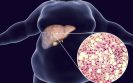
Scientists could help match cancer patients with no other treatment options to clinical trials with experimental medicines, by analysing the genetic faults in a sample of their blood, according to research published in Nature Medicine.

The researchers, funded by Cancer Research UK, The Christie Charity, AstraZeneca and the NIHR Manchester Biomedical Research Centre (BRC), demonstrated in their feasibility study that a blood test can be carried out and analysed in a timeframe that can help clinicians select a matched, targeted treatment.

Currently, enrolment to trials depends on a patient’s type of cancer or genetic data obtained from an invasive tumour biopsy, which is often months or years old and may not represent a patient’s current disease due to their tumours’ evolutionary changes over time.

Scientists from the Cancer Research UK Manchester Institute at The University of Manchester, showed that a small volume of blood can contain up-to-date genetic information about a patient’s cancer to inform treatment choices. In this feasibility study of the first 100 patients, 11 were enrolled onto an available and molecularly matched clinical trial.

Dr Matthew Krebs, the lead clinician of the study from The University of Manchester and The Christie Hospital NHS Foundation Trust, said: “This study is bringing clinicians and scientists together to develop a new approach to treating patients with advanced cancers.

Historically, patients who have exhausted other options but are still reasonably well might access a clinical trial based on their cancer type, but without that new therapy being targeted to their tumour’s particular genetic profile. Now, that paradigm is shifting toward personalised medicine. By understanding the genetic faults underpinning a patient’s cancer from a blood test, as demonstrated in this study, this raises the hope of matching more patients to a specific targeted clinical trial treatment with better chance of benefit.”

In the first of the two-part trial, called TARGET, the researchers were able to collect, process and analyse blood samples from 100 patients in the Manchester area.

Professor Caroline Dive, the laboratory lead author of the study from the Cancer Research UK Manchester Institute, said: “Now that we have demonstrated the feasibility of matching clinical trials for patients who have not responded to previous treatments by analysing the tumour DNA in their blood, we are working to improve our blood testing approach. We are making the test more sensitive and adding new elements to it in order to understand more about a patient’s disease. We are also taking several blood samples over time to see if a faulty gene(s) is disappearing with treatment, or if there is emergence of a new genetic fault that could lead to treatment resistance. This would allow us to stop a failing treatment and consider new options to stay a step ahead of the disease.”

The authors caution that while this study is promising, not every patient will have identifiable and ‘druggable’ faulty genes in their blood, nor will every patient have the opportunity to receive a treatment tailored to their cancer.

The researchers now hope the second part of TARGET, which is already underway, will show how often the blood test is successful at matching patients to early phase clinical trials and the impact this has on their overall survival. There is also an option of referring patients to other clinical trial sites, if suitable matched trials are available in other parts of the country.

Getting closer to finding out why the immune system attacks itself

15th April 2019

A comprehensive profile of B cells in rheumatoid arthritis (RA), a first of its kind study, has been published in Arthritis & Rheumatology.1 
 
To the best of our knowledge, this is the first study to conduct whole transcriptome profiling of antigen-specific B cells in any human autoimmune disorder,” said Navin Varadarajan, a researcher at the University of Houston, whose results portray B cells not merely as autoantibody producers, but also as a source of diverse molecules that can influence proliferation, differentiation and activation of other pathogenic cell types.
 
We anticipate that these data will serve as a foundational data set for investigating multiple hypotheses on the roles of B cells in RA and other autoimmune disorders, and will enable drug discovery,” said Varadarajan.
 
For every new pathogen encountered, a small subset of B cells activates to make an antibody that specifically recognises that particular pathogenic protein. Every person has between 10-100 million unique B cells, each capable of making its own antibody. In autoimmune RA, these antibodies – that are supposed to fight foreign invaders – attack the body’s own proteins and are thus called autoantibodies.
 
We wanted to understand if there’s anything special about this class of white blood cells, the autoreactive B cells that make autoantibodies, that would make them fight against healthy proteins,” said Varadarajan.
 
Fewer than one in 1000 B cells are autoreactive, so to find which one is the culprit, Varadarajan’s postdoctoral researcher Ankit Mahendra designed a method to reliably identify and isolate the population, then used RNA sequencing to study all of the RNA being made by each cell.
 
A number of pathways associated with inflammation and protein modification, known to be amplified in rheumatoid arthritis, were found. At the molecular level, the team found two specific differences in the B cells of RA patients – the inclusion of the protein interleukin 15 receptor subunit alpha (IL-15Rα) and a high amount of the amphiregulin molecule, which can signal adjacent cells. Each was validated at the protein level in independent cohorts of RA patients and prioritised for further studies.
 
We think that protein allows them to become bad actors,” said Varadarajan. “People have been targeting this pathway for quite some time. This now sheds new light on these bad guys in the progression of this disease and how to target it.”
 
The team is the first to show that B cells make amphiregulin. Amphiregulin sits in a well-studied pathway, the epidermal growth factor receptor pathway, and so the next step will be to determine if inhibiting the pathway impacts the B cells.
 
Reference
  1. Mahendra A et al. Beyond autoantibodies: Biologic roles of human autoreactive B cells in rheumatoid arthritis revealed by RNA‐sequencing. Arthritis & Rheumatology 2019;71(4):529 DOI: 10.1002/art.40772

Organisations join forces to create global alliance against antibiotic resistance

The US-based Antibacterial Resistance Leadership Group (ARLG), part of the Duke Clinical Research Institute (DCRI) and the University Medical Center (UMC) Utrecht, the managing entity of the COMBACTE (Combatting Bacterial Resistance in Europe) consortium, will work together to solidify a comprehensive global community to combat the threat of antibiotic resistance around the world.
 
This collaboration is expected to take several forms, including joint design and implementation of clinical research, working meetings at scientific conferences like the European Congress of Clinical Microbiology & Infectious Diseases (ECCMID), and IDWeek, cross-entity working groups with diverse functional group participation, clinical trial innovations, data and protocol exchanges, and, contractual, regulatory, and systems harmonisation.
 
We at the ARLG have long admired the work being done by COMBACTE to increase the efficacy of antimicrobial drug development,” said Vance Fowler, MD, an investigator at the DCRI and co-principal investigator of the ARLG. “Combining our efforts will allow us to maximise the work we both do to stop the advancement of antibacterial resistance.”
 
Both organisations have been working toward the same mission since each launched in 2013, and now, the two groups will share their work to increase synergy and avoid duplicative efforts in clinical research.
 
We have already made efforts to expand our reach across Europe, where an increasing number of people suffer from infections caused by antibiotic-resistant bacteria,” said Marc Bonten, MD, coordinator of COMBACTE and a professor at UMC Utrecht. “But by collaborating and sharing our progress with the ARLG, we can make a truly global impact beyond this population — an important consideration in a globally connected era in which epidemics travel across oceans quickly.”
 
As part of the agreement, the ARLG will have the opportunity to lead and coordinate U.S.-based studies for all clinical research initiated by COMBACTE, while COMBACTE will have the opportunity to lead and coordinate ARLG-initiated projects in Europe.
 
Earlier this year the European Clinical Research Alliance on Infectious Diseases (ECRAID) was formed, merging COMBACTE’s more than 850 clinical trial sites and 650 laboratories with the network of primary care sites coordinated by The Platform for European Preparedness Against (Re-)Emerging Epidemics (PREPARE) to form a European-wide sustainable clinical research organisation for infectious diseases and antimicrobial resistance. The ARLG, with established collaborations in 19 countries, will help to expand these efforts outside of Europe. The alliance will enable innovative, flexible, and adaptive collaboration between the DCRI and UMC Utrecht.
 
This is an exciting year for PREPARE to expand its work in reducing a serious public health threat,” said Herman Goossens, MD, coordinator of PREPARE and professor at University of Antwerp and UMC Utrecht. “First, we joined forces with COMBACTE by forming ECRAID, and now, we welcome a partnership with the ARLG, which will help all three organisations leverage a wider set of resources and a deeper pool of expertise.”

Expert analysis: The importance of confirming allergic triggers in rhinitis

Rhinitis is an inflammatory disorder of the nasal mucosa clinically defined by two or more symptoms of nasal itching, sneezing, anterior or posterior rhinorrhoea and nasal blockage.1

Rhinitis is considered chronic when the symptoms are present for at least one hour daily and last longer than two weeks.2 The lifetime prevalence rates of acute rhinitis and chronic rhinitis are 100% and >20%, respectively, which explains the considerable financial burden that the condition imposes to health care systems, in terms of consultations and medications prescribed.3

Moreover, chronic rhinitis negatively affects work and school performance and has been related to learning disabilities in children,4 and to an impairment in quality of life greater than that of chronic conditions such as arterial hypertension.5

Chronic rhinitis is often associated to other inflammatory disorders of the mucous membranes such as sinusitis, conjunctivitis and asthma, which further amplifies its impact.6 Nevertheless, the condition has been historically trivialised and has been regarded as a relevant health problem only in recent years.

Classifications

Several classifications have been proposed for chronic rhinitis based on different parameters such as pathophysiology, frequency and pattern of symptoms or trigger(s) eliciting rhinitis.2

One easy classification divides the disorder into allergic rhinitis (AR) and non-allergic rhinitis (NAR). AR is the most frequent form of chronic rhinitis,7 and constitutes a relatively homogenous phenotype with known pathophysiology defined by IgE-sensitisation to environmental allergens.6

Conversely, NAR comprises a highly heterogeneous group of diseases where immune-mediated inflammation is not always apparent.8 NAR patients are defined by rhinitis symptoms and negativity of classical IgE-sensitisation tests, namely the skin prick test (SPT) and serum allergen-specific IgE7 (Figure 1).

Figure 1: Diagnostic biomarkers

Figure 1: Diagnostic biomarkers in the allergic phenotypes of rhinitis. Allergic rhinitis (AR) is characterised by the positivity of skin prick (SPT) test and serum specific (s)IgE. These two biomarkers are sufficient for diagnosis in many cases of AR. In AR individuals, the clinical relevance of an IgE-sensitisation can be accurately established by a nasal allergen challenge (NAC). The basophil activation test (BAT) and the nasal sIgE are positive in most cases of AR, even though they do not use to be necessary for diagnostic purposes. By definition local allergic rhinitis (LAR) patients test negative for both SPT and serum sIgE. The diagnosis of LAR is routinely established by the positivity of the NAC. Some LAR individuals have detectable nasal sIgE or positive BAT responses, and these tests can assist to reach the diagnosis.

Symptoms of AR are driven by re-exposure to seasonal or perennial allergens in IgE-sensitised individuals,1 and AR patients are by definition positive for at least one of the two tests to measure IgE sensitisation: SPT and/or serum allergen-specific IgE.6

Nevertheless, a significant proportion of healthy subjects also display positivity for either test, demonstrating that the correlation of symptoms with allergen exposure is crucial for the interpretation of IgE-sensitisation tests.9

A nasal allergen challenge (NAC) can help determine the clinical relevance of IgE-sensitisation in this setting10 (Figure 2).

Interestingly, some patients with seasonal or perennial rhinitis symptoms display positive NAC with negative SPT and serum allergen-specific IgE. Our group and others have studied this disease phenotype, which is usually termed local allergic rhinitis (LAR).11,12

This disorder does not properly fit into the above mentioned AR/NAR dichotomy. Similar to AR, LAR patients have a type 2-dominated nasal inflammation, including activation of resident mast cells and recruitment and activation of eosinophils.13,14

Moreover, LAR patients share many clinical features and in vitrofindings with AR individuals, importantly the positivity of the NAC,15 and the presence of allergen-specific IgE in the nasal secretions.16

Epidemiological studies have demonstrated that LAR is a moderate-to-severe condition that might affect up to 25% of non-atopic patients with rhinitis, and that tends to worsen over time,17 with associated onset of asthma and conjunctivitis.18 LAR is not an initial state of AR, as studies from our group show that the long-term conversion rate to systemic atopy is comparable between LAR subjects and the general population.17

Figure 2: Diagnostic algorithm of the allergic forms of rhinitis

Figure 2: Diagnostic algorithm of the allergic forms of rhinitis. BAT: basophil activation test; NAC: nasal allergen challenge; NsIgE: allergen-specific IgE in the nasal secretions.

Identification of triggers is crucial

Identification of the allergic triggers of rhinitis is crucial in order to implement adequate avoidance measures in both atopic and non-atopic patients. Moreover, this identification can help administer specific therapies, such as allergen immunotherapy (AIT). Several controlled trials from different groups have demonstrated the capacity of AIT (with house dust mites, grass and tree pollens) to control the symptoms, reduce the need for rescue medication, improve quality of life and increase the nasal tolerance to the allergen in LAR individuals19–21 (Table 1).

Table 1: Studies that have investigated the performance of AIT in LAR patients

Table 1: Studies that have investigated the performance of AIT in LAR patients. In all cases similar results and conclusions were obtained: AIT is able to control the symptoms of LAR patients, to reduce the need for rescue medication, to improve quality of life, and to increase the amount of allergen tolerated in the NAC. The red square highlights the three randomised, double-blind, placebo-controlled clinical trials (RDBPCCT) published to date.

In AR patients, AIT is able to prevent the progression of the disease, especially the onset of asthma.22 It is currently under investigation whether AIT has the same capacity in the LAR phenotype. Importantly, there is a rapid evolution towards the clinical worsening during the first five years after LAR onset,17 implying that this initial period represents a window of opportunity to establish specific therapies aiming to prevent the progression of the disease.

All the above mentioned aspects highlight the relevance of identifying the allergic triggers of nasal reactivity, regardless of the atopic status of the rhinitis patient. In this regard, the NAC is the key tool for the diagnosis of LAR and it can confirm the clinical relevance of IgE-sensitisations in AR individuals.23

Moreover, the NAC can help design the composition of AIT in patients with multiple sensitisations, or monitor its effect.23 The NAC displays the optimal features for an in vivo test, as it is very safe and reproducible.24 The NAC for LAR diagnosis significantly outperforms the detection of nasal allergen-specific IgE and the basophil activation test in terms of both sensitivity and specificity.23,24 

Conclusions

In summary, there is a significant proportion of non-atopic rhinitis patients who display nasal reactivity to environmental allergens. Therefore, the inclusion of the NAC in the diagnostic algorithm is necessary to accurately identify the allergic triggers of rhinitis. This identification is crucial for the early prescription of specific therapies such as AIT which has the potential to prevent the progression of the disease, including the onset of asthma.

Authors

Ibon Eguiluz-Gracia MD PhD

Adriana Ariza PhD

Alba Rodríguez-Nogales PhD

Carmen Rondon MD PhD

References

1 Greiner AN et al. Allergic rhinitis. Lancet 2011;378(9809):2112–22.

2 Papadopoulos NG, Guibas GV. Rhinitis subtypes, endotypes, and definitions. Immunol Allergy Clin NAm 2016;36(2):215–33.

3 Canonica GW et al. A survey of the burden of allergic rhinitis in Europe. Allergy. 2007;62 Suppl 85:17–25.

4 Muraro A et al. The management of the allergic child at school: EAACI/GA2LEN Task Force on the allergic child at school. Allergy 2010;65(6):681–9.

5 de la Hoz Caballer B et al. Allergic rhinitis and its impact on work productivity in primary care practice and a comparison with other common diseases: the Cross-sectional study to evAluate work Productivity in allergic Rhinitis compared with other common dIseases (CAPRI) study. Am J Rhinology Allergy 2012;26(5):390–4.

6 Bousquet J et al. Allergic rhinitis and its impact on asthma. J Allergy Clin Immunol 2001;108(5 Suppl):S147–334.

7 Dykewicz MS, Hamilos DL. Rhinitis and sinusitis. J Allergy Clin Immunol 2010;125(2 Suppl 2):S103–15.

8 Rondon C et al. Nonallergic rhinitis and lower airway disease. Allergy 2017;72(1):24–34.

9 Roberts G et al. A new framework for the interpretation of IgE sensitization tests. Allergy 2016;71(11):1540–51.

10 Dordal MT et al. Allergen-specific nasal provocation testing: review by the rhinoconjunctivitis committee of the Spanish Society of Allergy and Clinical Immunology. J Invest Allergol Clin Immunol 2011;21(1):1–12; quiz follow Epub 2011/03/05.

11 Krajewska-Wojtys A, Jarzab J, Gawlik R, Bozek A. Local allergic rhinitis to pollens is underdiagnosed in young patients. Am J Rhinol Allergy 2016;30(6):198–201.

12 Rondon C, Canto G, Blanca M. Local allergic rhinitis: a new entity, characterization and further studies. Curr Opin Allergy Immunol 2010;10(1):1–7.

13 Rondon C et al. Nasal inflammatory mediators and specific IgE production after nasal challenge with grass pollen in local allergic rhinitis. J Allergy Clin Immunol 2009;124(5):1005–11 e1.

14 Rondon C et al. Local IgE production and positive nasal provocation test in patients with persistent nonallergic rhinitis. J Allergy Clin Immunol 2007;119(4):899–905.

15 Rondon C et al. Nasal allergen provocation test with multiple aeroallergens detects polysensitization in local allergic rhinitis. J Allergy Clin Immunol 2011;128(6):1192–7.

16 Campo P et al. Local IgE in non-allergic rhinitis. Clin Exp Allergy Immunol 2015;45(5):872–81.

17 Rondon C et al. Follow-up study in local allergic rhinitis shows a consistent entity not evolving to systemic allergic rhinitis. J Allergy Clin Immunol 2014;133(4):1026–31.

18 Rondon C et al. Prevalence and clinical relevance of local allergic rhinitis. Allergy 2012;67(10):1282–8.

19 Rondon C et al. Efficacy and safety of D. pteronyssinus immunotherapy in local allergic rhinitis: a double-blind placebo-controlled clinical trial. Allergy 2016 Jul;71(7):1057–61.

20 Rondon C et al. Specific immunotherapy in local allergic rhinitis: A randomized, double-blind placebo-controlled trial with Phleum pratense subcutaneous allergen immunotherapy. Allergy 2018 Apr;73(4):905–15.

21 Bożek A, Kołodziejczyk K, Jarząb J. Efficacy and safety of birch pollen immunotherapy for local allergic rhinitis. Ann Allergy Asthma Immunol 2018;120(1):53–8.

22 Durham SR et al. SQ-standardized sublingual grass immunotherapy: confirmation of disease modification 2 years after 3 years of treatment in a randomized trial. J Allergy Clin Immunol 2012;129(3):717–25.e5.

23 Augé J et al. EAACI Position paper on the standardization of nasal allergen challenges. Allergy 2018;73(8):1597–608.

24 Eguiluz-Gracia I et al. Safety and reproducibility of nasal allergen challenge. Allergy 2019; in press.

High-tech tissue study reveals which cells drive a painful food allergy

12th April 2019

An eight-year hunt for the cells that drive the extreme childhood food allergy eosinophilic esophagitis (EoE) has identified a potential new way to treat the disease while also raising questions about a dietary supplement often taken to reduce bowel inflammation.
 
The study, led by experts at Cincinnati Children’s Hospital Medical Center, is posted online in the Journal of Clinical Investigation1 and will be published in print in May. 
 
Reported cases of EoE have risen sharply in the past two decades, now affecting as many as one of every 2000 people. This lifelong allergic disease occurs when too many eosinophils cause inflammation that makes swallowing and eating painful. EoE can be so serious that some people depend upon special liquid formulas to eat.
 
In this study, Wen and Rothenberg developed and perfected a 10- to 12-hour process that isolates and analyses individual living cells from human tissue samples about the size of three sesame seeds.
 
This is actually a substantial advance for the field,” Rothenberg says. “Prior work has focused on blood cells, but not the specific memory immune cells in tissue that directly respond to food allergens. We uncovered the unknown properties of these cells.”
 
Their key findings:
  1. Eight types of immune system T cells were found in the oesophageal tissue. That’s more than had been previously expected.
  2. Two cell types (T7 and T8) show up in higher numbers in the diseased tissue. The study finds that one of these two cell types responds to allergens by cranking out massive amounts of inflammation-causing type 2 cytokines.
  3. One of these cell types (T8) is nearly non-existent in healthy tissue, making it especially worth targeting for treatment. “These cells are bad actors that should not be in this type of tissue. Inhibiting these cells is now a central goal of our research,” Wen says.
  4. So how did these bad players get there? Studying this question led the co-authors to a surprising finding. One of the overactive genes that helps trigger the inflammation response – a gene called FFAR3 – also encodes a receptor for the dietary supplement butyrate.
Beyond EoE, the process used in this study could be applied in other, more-common conditions to find small populations of cellular culprits hiding within tissues. The study co-authors say the process could be useful in studying asthma, atopic dermatitis, allergic rhinitis, and other diseases associated with type 2 cytokines.
 
Reference
  1. Wen T et al. Single-cell RNA sequencing identifies inflammatory tissue T cells in eosinophilic esophagitis. J Clin Invest 2019;DOI: 10.1172/JCI125917

First results of a major three-year project to analyse airborne grass pollen

A study published in Nature Ecology & Evolution has shown that it is not just the overall ‘load’ of grass pollen in the air that could cause those particularly bad days for asthma and hay fever sufferers.1 Days that see increased asthma attacks or intense hay fever could be related to the release of pollen from particular grass species.
 
Current pollen ‘counts’ and forecasts assess the whole load of pollen in the air, and, while scientists can distinguish between the pollen created by individual tree and weed species, it has proven virtually impossible for the current forecast methods to visually identify different grass pollens.
 
Metabarcoding is a technique which enables scientists to automatically identify any fragments of material caught in a sample of air, water or soil, by recognising and matching its unique DNA ‘barcode’.
 
For the first time, grass pollens collected over the course of one allergy season have been analysed using this high-tech method. This has enabled the team to start investigating links between certain pollen types and those days on which plant allergy sufferers and people with asthma are most affected.
 
Professor Simon Creer, of Bangor University, who is leading the research, explains: “I’m a hay fever sufferer myself, and I know that on some days, despite a high pollen forecast, I can be less affected than on other days when the forecast appears to be lower. This led me and others to wonder whether it’s the high load of pollen alone that causes the problem, or whether the different grass pollens cause different levels of reaction.”
 
Dr Georgina Brennan, from Bangor University who analysed the aerial pollen “environmental DNA” with Dr Caitlin Potter from the University of Aberystwyth and the National Botanic Garden Wales, added:”Bringing a range of specialists together has enabled us to find initial answers. Our task is now to develop a clearer picture of where the pollen comes from, how it moves through the air and how different types of pollen can be linked to allergies.”
 
Dr Ben Wheeler of University of Exeter said: “We are now investigating datasets on hospital admissions and GP prescriptions for certain pharmaceutical products to identify correlations between healthcare data and increases in particular grass pollens. With these new insights into pollen characterization, we are focusing on future implications for pollen warnings and self-care strategies.”
 
Dr Rachel McInnes from the UK Met Office added: “Leading on from this new environment DNA research, we are currently developing maps of where these species of allergenic grasses are located in the UK. When combined with aerial modelling approaches being developed with Prof. Carsten Skjøth at the University of Worcester, the approaches could be used to improve our pollen forecast in the future.”
 
Reference
  1. Brennan GL et al. Temperate airborne grass pollen defined by spatio-temporal shifts in community composition. Nature Ecol Evolution 2019;DOI: 10.1038/s41559-019-0849-7

Researchers develop first functional targeted inhibitors of peanut allergens

In a study published in the Proceedings of the National Academy of Sciences, researchers at the University of Notre Dame in the US have effectively prevented the binding of peanut allergens with IgE to suppress the allergic reaction to peanuts using a first-in-class design of allergen-specific inhibitors.
 
The success of this study is exciting because it opens the door to establishing an entirely new class of allergy therapeutics,” said Basar Bilgicer, associate professor in the Department of Chemical and Biomolecular Engineering and affiliate of Advanced Diagnostics and Therapeutics at Notre Dame, who led the study. “We now have the first functional example of selective IgE inhibition to a food allergen, which we haven’t had before.”
 
For Bilgicer, the challenge was developing an inhibitor that is designed to specifically inhibit the binding of IgE and the allergen proteins without interfering with any other immune system functions. Food allergen-specific inhibitors would stop degranulation, the primary driver of life-threatening allergic reactions, without putting the patients’ immune system or their health at risk.
 
To achieve their goal, Bilgicer and his team used nanoparticles, called nanoallergens, to screen and identify the key binding sites on peanut proteins that patient IgE antibodies recognise by studying samples from a small population of patients with severe allergies to peanuts. That was significant, Bilgicer said, because “it seems only a few sites seem to be exceptionally critical in driving the allergic response.” Once identified, Bilgicer’s team synthesised a specialised inhibitor – the covalent heterobivalent inhibitor (cHBI) – to prevent IgE from binding to the peanut protein. In a study of 16 patient samples with severe peanut allergies, the cHBIs were successful in inhibiting an allergic response in up to 90% of all samples.
 
The study presents a compelling case for further development and assessment of cHBI as a viable strategy for treating peanut allergies.
 
Reference
  1. Deak PE et al. Designer covalent heterobivalent inhibitors prevent IgE-dependent responses to peanut allergen. PNAS 2019;April 8:DOI: 10.1073/pnas.1820417116

x