This website is intended for healthcare professionals only.

Hospital Healthcare Europe
Hospital Pharmacy Europe     Newsletter       

Press Releases

Take a look at a selection of our recent media coverage:

Record number of patients benefit as clinical trial participants soar across England

20th May 2019

The past 12 months has shown a positive rise in people benefiting from clinical research in England, as numbers have reached record highs with over 870,000 participants involved in clinical research studies supported by the National Institute for Health Research (NIHR) over the last year.
 
This high number takes significant steps towards the NHS Long Term Plans goal to reach one million people in trials by 2023/2024, which is part of the Government’s overall strategy to improve care, treatment and NHS services in England.
 
Health research for children has recruited the most participants this year with 81,892 people taking part. This was followed by studies delivered in primary care settings (78,533). Childbirth and sexual health research recruited the third highest number of participants at over 74,128 – up 20% from last year’s figures.
 
The latest figures from NIHR show that patients now have more opportunity than ever before to take part in clinical research and potentially benefit from new drugs and treatments. A survey of over 8,500 participants over the last year showed that 90% reported a good experience of taking part in research.
 
Dr Jonathan Sheffield OBE, Chief Executive of the NIHR Clinical Research Network says “We are delighted that this year alone, hundreds of thousands of people across the country have given their time to improve healthcare for others. Without their commitment, vital health research that changes lives simply could not happen”.
 
The benefits that clinical research bring to society are profound. People who take part in studies can gain access to cutting edge, innovative new treatments. While NHS trusts and health and social care patients also benefit significantly, with evidence and innovations identified through research pivotal to the development of new types of care and treatment – ultimately leading to the prevention of ill health, earlier diagnosis, faster recovery and better outcomes.”
 
Baroness Blackwood, Parliamentary Under Secretary of State at the Department of Health and Social Care said: “From the eradication of smallpox and the discovery of penicillin, the UK has a strong track record of public health successes which have saved countless lives. All of our successes to date would have been impossible without world-leading research and the selfless volunteers who take part in clinical trials.” 

New strategy of reprogramming regulatory T cells may improve cancer therapies

17th May 2019

A new study from the Center for Immunology and Inflammatory Diseases (CIID) at Massachusetts General Hospital (MGH) describes a method of reprogramming the regulatory T cells that usually suppress immune responses into inflammatory cells that not only permit but also intensify an antitumour immune response.1 
 
Many patients’ tumours do not respond to immune therapies – such as immune checkpoint blockade – because of a lack of pre-existing inflammation that is required for those therapies to work,” says Thorsten Mempel, MD, PhD, of the MGH CIID, senior author of the paper. “Our study shows that reprogrammed Treg cells provide exactly the type of inflammation that is lacking. Indeed, we found in mice that reprogramming tumour-infiltrating Treg cells to secrete inflammatory cytokines makes previously unresponsive tumours highly sensitive to PD-1 blockade.”
 
The study focused on the CBM complex – a large protein cluster within immune cells that helps regulate their activation, proliferation and function. Recent research has revealed a critical role for the CBM complex in lymphocyte function, and since deleting one of three key proteins, called CARMA1, is already known to reduce the function of effector T cells, the team examined the effects of CARMA1 deletion on Treg cells.
 
Their experiments revealed that targeting the CBM complex – either by deleting one or both copies of the CARMA1 gene in Treg cells or by treating tumour-bearing mice with a drug that inhibits MALT1, another component of the complex – caused Treg cells to secrete the immunostimulatory cytokine interferon gamma in tumour tissue alone. The ability to selectively modulate the function of Treg in tumours can avoid the risk of autoimmune disease that would result from systemic Treg depletion.
 
CBM targeting led to inflammation of tumour tissue and increased infiltration by cytotoxic CD8 T cells and natural killer cells. But it only reduced the rate of tumour growth in mouse models of melanoma and colon cancer because the activity of those immune cells was still limited by the immune checkpoint protein PD-1. However, blocking the activity of PD-1 with antibodies led to elimination of tumours that had been inflamed by anti-CBM treatment.
 
Reference
  1. Di Pilato M et al. Targeting the CBM complex causes Treg cells to prime tumours for immune checkpoint therapy. Nature 2019; DOI: 10.1038/s41586-019-1215-2

New technology to revolutionise drug development

The search for new drugs to combat diseases more effectively could be revolutionised through a new £30 million electron microscopy project.
 
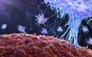
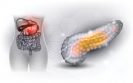
The technology aims to create 3D images of cells at very high resolution to transform our understanding of diseases such as cancer and revolutionise how new medicines are designed.
 
The project based at Harwell, in Oxfordshire, will be supported with £3 million from the government’s Office for Life Sciences and led by the global life sciences company Thermo Fisher Scientific and the Rosalind Franklin Institute, which is dedicated to bringing about transformative changes through research and technology development. 
 
Professor James Naismith, lead researcher and Director of the Rosalind Franklin Institute, said: “Around 80% of new drugs fail when they reach clinical trials because we don’t fully understand their effects within a living organism.
 
We’re currently able to identify genes and proteins that play a role in diseases but can only study these in detail in isolation, not as part of a whole cell or group of cells. This new technology would be the first to allow us to do that, so we can see the full effects of a drug and identify at a much earlier stage what will work and what will not.
 
Drug development has never been slower or higher cost than it is now. We need to have a breakthrough in the physical science that supports drug discovery to change that.
 
The project was announced on 17 May 2019 as work began on the Rosalind Franklin Institute’s central hub building on the Harwell Campus near Oxford. The building will create a specialist environment for sensitive scientific equipment that will be unique in the world. It is set to house 200 researchers and will open in late 2020.
 
The new imaging technique will be based on a technology called cryogenic electron tomography (cryo-ET), which builds up a 3D image from multiple 2D images of samples which have been flash frozen at temperatures below -180oC. 
 
Although cryo-ET already exists, it can currently only handle small samples, such as parts of cells, and often requires in-house expertise.
 
Mike Shafer, president of materials and structural analysis at Thermo Fisher Scientific said: “Our company’s mission is to enable our customers to make the world healthier, cleaner and safer, and our collaboration with the Rosalind Franklin Institute, Diamond Light Source and others to develop a more efficient cryo-tomography solution, maps precisely to that vision
 
Our combined goal is to make this technique widespread so scientists and researchers in academic institutions and the biopharmaceutical industry can better understand disease mechanisms at the cellular level, leading to faster drug discovery and cures for neurodegenerative diseases, cancers and other debilitating illnesses.”
 
The new, five-year research project will rapidly speed up the technique to process much larger samples including patient biopsies, increase workflow automation and standardise post-processing data.
 
The long-term aim is to bring the technology closer to the clinic so it can be used in both research hospitals and laboratories.
 
The challenges around the project are formidable. It will require the invention of new techniques for preparing and handling samples of human tissue that are less than a hundredth of the thickness of human hair.
 
It will involve the design of new electron microscopy techniques to speed up the imaging process and collect the huge amount of data created. New software and machine learning technology will need to be developed to process this data in order to create and interpret the 3D images.
 
To tackle the challenges, the project brings together an impressive array of expertise from the different research partners. Thermo Fisher Scientific is a global leader in scientific instrumentation; Diamond Light Source, the UK’s national synchrotron facility includes one of the largest industrial cryo-electron microscopy sites in the world; and the Rosalind Franklin Institute, funded through UK Research and Innovation, combines scientific expertise from ten of the UK’s leading universities.
 
Andrew Bourne, Deputy Director at UKRI’s Engineering and Physical Sciences Research Council (EPSRC), said: “Today we are building for the future of the UK’s research in life sciences and the development of new drugs to benefit us all. This new project is a great example of collaboration between industry, the academic community, and government funding bodies. It is also fantastic to see work beginning on the hub building for the Rosalind Franklin Institute, which has been delivered and managed by UKRI’s Engineering and Physical Sciences Research Council. The Institute’s work will continue the longstanding relationship between research discovery in engineering and the physical sciences and advances in medicine. It will help strengthen the UK’s position in the global research landscape and develop new medical treatments and technologies in the life sciences.” 
 
Prof Peijun Zhang Director of the electron Bio-Imaging Centre (eBIC), the UK’s national cryo-EM centre, and part of Diamond, adds: “eBIC is keen to help push forward this development of in situ biological imaging. We provide existing state of the art instrumentation and expertise to enable cutting edge cell biology science, with access for the UK and worldwide science community using the synchrotron beamline model that changed the face of macromolecular crystallography. We look forward to feeding the new developments through to users as quickly as possible
 
The collaboration represents a major investment and is testament to the scientific strengths and long history of the UK in biological imaging and structural biology. The project is part-funded by the Department for Business, Enterprise and Innovation through the Office for Life Sciences as part of the government’s Life Sciences Industrial Strategy.

Liquid biopsy could identify cancer patients at risk of metastatic disease

Researchers at The Royal Marsden NHS Foundation Trust in the UK say ctDNA, a form of liquid biopsy, may be an accurate technique to monitor treatment response in patients with locally advanced rectal cancer, allowing treatment to be adapted or changed earlier to try to prevent the development of metastatic disease.

Researchers analysed liquid biopsies from 47 patients at The Royal Marsden who had localised rectal cancer – i.e. cancer that had not already spread to other body parts. They took blood samples before, during and after patients had completed treatment with combined chemotherapy and radiotherapy (CRT), and after surgery. They were able to detect ctDNA in 74% of patients pre-treatment, 21% of patients mid-way through CRT, 21% after CRT and 13% after surgery. The ctDNA results at the end of CRT were associated with tumour response to CRT as shown on MRI scans.

With a median follow up of just over two years, they found ctDNA results to be consistent with occurrences of cancer spreading outside of the rectum; patients with ctDNA persisting throughout their treatment, were more likely to develop metastatic disease sooner.

Lead author Dr Shelize Khakoo, Medical Oncologist at The Royal Marsden NHS Foundation Trust said: “We know patients respond quite differently to standard treatment – some will do really well, and have what we call a complete pathological response. For others, the cancer may spread during treatment.

“If we can predict early on who will go on to develop metastatic disease, we might be able to tailor treatment by making it more intense or trying an alternative.”

Co-lead author Professor David Cunningham OBE, Consultant Medical Oncologist at The Royal Marsden NHS Foundation Trust said: ‘These results suggest that liquid biopsies offer us an accurate method of establishing the cancer’s activity throughout the body.

“Importantly what this study showed, which has not yet been explored, is that persistence of ctDNA mid-way through treatment could be an early indicator of the cancer’s potential to spread. Using this measure, along with MRI scans, we can offer a more personalised treatment approach for patients.”

Is FMT helpful in children with C. difficile?

A study in the journal Clinical Gastroenterology and Hepatology, the largest faecal microbiota transplant (FMT) study in children to date, found it both safe and effective and also identifies predictors of success.
Diarrhoeal disease from C. difficile on the rise among children. One population-based study found a 12.5-fold increase in incidence from 1991 to 2009. For reasons that aren’t clear, C. difficile is more frequently striking children without such risk factors as hospitalisation or antibiotic exposure.
“Our results are quite exciting, given that recurrent C. diff is debilitating and increasing in children, not just those with known risk factors but previously healthy children as well,” says gastroenterologist Stacy A. Kahn, MD, at Boston Children’s Hospital. Kahn led the study with Maribeth Nicholson, MD, MPH, at Vanderbilt University Medical Center and Richard Kellermayer, MD, PhD at Texas Children’s Hospital.
The team retrospectively studied 372 patients with C. difficile, age 11 months to 23 years, who had FMT at one of 18 paediatric centres across the United States. FMT was administered in various ways, including colonoscopy, nasogastric tube, frozen capsules, or enemas.
Two-month outcomes were available for 335 patients. Of these, 81% had no recurrence of C. difficile infection after a single treatment. Some of the remaining patients had a second round of FMT; about half of them saw no C. difficile recurrence, increasing the overall success rate to 87%.
Success was 2.7 times more likely when FMT came in the form of fresh versus previously frozen stool, and 2.4 times more likely when patients received the stool via colonoscopy versus other methods. Patients without a feeding tube (itself considered a risk factor for C. difficile) were twice as likely to respond, and those with one fewer prior C. difficile infection had a 20 percent greater chance of success. Age did not appear to be factor.
FMT was also more likely to succeed in patients treated more recently, perhaps because of tighter protocols around donor selection and treatment.
“The success rates we found are similar to what is seen in adults, but seem to be associated with fewer complications,” says Kahn. “We don’t understand why colonoscopic delivery, fresh stool and more recent FMT treatment were associated with higher success rates. These questions provide the foundation for future FMT research in children.”
As for safety, 6% of the patients had FMT-related adverse events; most were mild and included diarrhoea, vomiting, and bloating. Of the roughly one-third of patients who also had inflammatory bowel disease, 2.5% had a severe flare of their illness requiring them to be hospitalised. But it wasn’t clear that had anything to do with the FMT.
Kahn notes that the study was limited by its retrospective design and relatively short period of follow-up. “We are using the data to help design future prospective, controlled studies,” she says. “We also need longitudinal studies to investigate the long-term impact and safety of transplanting the microbiome from one person to another. A lot more questions need to be answered.”
The group is also studying FMT for children with other forms of colitis, such as inflammatory bowel disease.
We want to tap into the therapeutic potential of FMT and begin to understand how we can use and manipulate the microbiome to treat diseases other than C. diff,” Kahn says.

Whole body MRI may help to detect spread of cancers more quickly

10th May 2019

Trials with people with newly-diagnosed colorectal and non-small cell lung cancer suggest that whole body MRI could reduce the time it takes to diagnose the stage of cancers.
 
The results are from two prospective trials with nearly 500 patients across 16 UK hospitals, published in The Lancet Gastroenterology & Hepatology and The Lancet Respiratory Medicine journals.1,2
 
Whole body MRI scans reduced the average time to determine the size of tumours and how much they had spread by five days for colorectal cancer patients and six days for lung cancer patients. The treatments decided upon were similar, since results from MRI were as accurate as from standard investigations, but the costs per patient were reduced by nearly a quarter in the case of colorectal cancer and were almost halved for lung cancer. More research is needed to determine how this affects outcomes for patients. 
 
Despite their accuracy and efficiency, the authors note that MRI scanners are not as widely available as other imaging technologies and are in high demand. In the trials, many of the hospitals were not able to find time on their MRI scanners, meaning that patients were examined in nearby hospitals. 
 
Our results, obtained in a real-world NHS setting, suggest that whole body MRI could be more suitable for routine clinical practice than the multiple imaging techniques recommended under current guidelines,” says lead author Professor Stuart Taylor from UCL, UK. “While demands on NHS MRI scanners is currently high, MRI can image the whole body in one-hour or less Adopting whole body MRI more widely could save rather than increase costs, as well as reducing the time before a patient’s treatment can begin.”
 
Appropriate treatment cannot be decided upon until the size of a tumour and the extent to which it has spread to nearby lymph nodes and other parts of the body has been determined. Standard NHS pathways often involve different imaging techniques – such as CT, PET-CT or focused MRI scans – which vary in accuracy in different organs. Several appointments and follow-up examinations can therefore be necessary. 
 
For the first time, the two new trials compare the diagnostic accuracy and efficiency of whole body MRI with the standard NHS pathways, which use a range of imaging techniques for assessing colorectal and lung cancers. The standard imaging tests recommended by the National Institute for Health and Care Excellence were undertaken as usual and the usual multi-disciplinary panel made a first treatment decision based on their results. Once this decision had been recorded, they considered images and reports from whole body MRI. If the latter highlighted a need for further tests, these were carried out. The panel were then able to say whether their first treatment decision would have different based on WB-MRI result. In the interests of patient care, the final decision was made based on results from all tests.
 
Patients were also followed up after 12 months to better evaluate the accuracy of whole body MRI compared with standard tests. For example, whether one approach was more sensitive than the other in detecting spread of the primary tumour to other parts of the body. Based on this data, the panel were able to retrospectively evaluate what the optimal treatment decision should have been. 
 
Sensitivity and specificity of diagnosis for whole body MRI did not differ from standard tests for both cancers. The use of whole body MRI reduced the time it took to complete diagnostic tests, from an average of 13 days to an average of 8 days in the colorectal cancer trial and from 19 days to 13 days in the lung cancer trial. Costs were reduced from an average of £285 to £216 in the colorectal cancer trial and from an average of £620 to £317 in the lung cancer trial. 
 
In the colorectal cancer trial, agreement with the final multi-disciplinary panel treatment decision based on standard investigations and whole body MRI was similar and high (95% and 96%, respectively), as were results for the lung cancer trial (99% for standard investigations, and 98% for whole body MRI). 
 
Eight of the 16 hospitals in the colorectal cancer trial and 11 of the 16 hospitals in the lung cancer trial did not have the infrastructure to perform whole body MRI. 
 
The authors note that their findings are specific to colorectal and non-small cell lung cancer and might not be relevant to tumours arising in other parts of the body. In addition, waiting times might not be representative of other UK hospitals or of hospitals in other countries. A further limitation of the lung cancer trial is that sensitivity in detecting the spread of cancers – including the development of secondary tumours and the spread to lymph nodes – was low using both current standard imaging techniques and whole body MRI. Further research is needed to improve the performance of non-invasive imaging.
 
Writing in a linked Comment, Professor Andreas Schreyer from Brandenburg Medical School, Germany, says of the colorectal cancer trial: “MRI has faced considerable backlash within the medical community due to relatively high costs and the problems involved in finding a timely slot for imaging because of the high demand for this method. This is why it is particularly important to think outside the box and look out for new medical pathways and paradigms and not to be driven by prejudices. It could be more efficient to adapt the known therapeutic concept of hitting hard and early to diagnostic imaging to improve medical outcomes and economic performance.” 
 
References
  1. www.thelancet.com/journals/langas/article/PIIS2468-1253(19)30056-1/fulltext
  2. www.thelancet.com/journals/lanres/article/PIIS2213-2600(19)30090-6/fulltext 

PICO wound care device receives NICE guidance

9th May 2019

Smith & Nephew welcomes the launch of new medical technologies guidance from the UK National Institute for Health and Care Excellence (NICE). 
 
In the guidance, NICE recommends that PICO Single Use Negative Pressure Wound Therapy System (sNPWT) should be considered as an option for closed surgical incisions in patients who are at high risk of surgical site infections (SSIs).1 Key patient risk factors include a high BMI, diabetes, renal insufficiency and smoking.2
 
NICE concluded that PICO sNPWT is associated with fewer SSIs and seromas compared with standard wound dressings across several types of surgery. Cost modelling suggests that compared with standard wound dressings, PICO sNPWT provides extra clinical benefits at similar overall cost to the NHS.1 For some types of surgery, PICO sNPWT is cost saving.1
 
When making its recommendations, NICE considered a review of 31 studies, 15 of which were randomised controlled trials.1 A supporting meta-analysis showed that PICO reduced the risk of SSIs by 63%, the risk of seroma by 77% and the risk of dehiscence by 30%.3 Significantly, it also showed there was an almost 2-day reduction in length of hospital stay, providing substantial cost savings and efficiency gains across the healthcare system.3
 
Surgical site complications are an increasing concern for healthcare providers and patients,” said Simon Fraser, President, Advanced Wound Management, Smith & Nephew. “NICE’s recognition of the proven impact PICO can make on both clinical outcomes and cost efficiencies will hopefully challenge existing standards of care around the world.”
 
The PICO sNPWT dressing includes a proprietary AIRLOCK™ Technology layer that uniformly and consistently delivers sufficient NPWT across a surgical incision and the surrounding zone of injury.4,5 This unique feature is designed to help reduce the risk of wound complications by reducing post-operative fluid6,7 and tension8 around a closed surgical incision, when compared with standard dressings. The combination of these actions helps reduce the risk of surgical wound dehiscence3 and SSIs,3 the two most common surgical site complications.
 
References
  1. NICE Medical Technology Guidance MTG43. PICO Negative Pressure Wound Dressings for closed surgical incisions. May 9th 2019
  2. World Union of Wound Healing Societies (WUWHS) Consensus Document. Closed surgical incision management: understanding the role of NPWT. Wounds International, 2016.
  3. Smith & Nephew. April 2019. Outcomes following PICO compared to conventional dressings when used prophylactically on closed surgical incisions: systematic literature review and meta-analysis. Report reference EO/AWM/PICO/004/v3
  4. Smith & Nephew October 2017. Project Opal PICO 7 System Stability Testing, Initial Time Point. Internal Report. DS/17/253/R.
  5. Malmsjö M et al. Biological Effects of a Disposable, Canisterless Negative Pressure Wound Therapy System. ePlasty. 2014;14.
  6. Karlakki SL, Hamad AK, Whittall C, et al. Incisional negative pressure wound therapy dressings (iNPWTd) in routine primary hip and knee arthroplasties: A randomised controlled trial. Bone Joint Res. 2016;5(8):328-337.
  7. Payne C, Edwards D. Application of the Single Use Negative Pressure Wound Therapy Device ( PICO ) on a Heterogeneous Group of Surgical and Traumatic Wounds. ePlasty. 2014:152-166.

Volanesorsen receives conditional marketing authorisation from the European Commission

Akcea Therapeutics and Ionis Pharmaceuticals have announced that Waylivra (volanesorsen) has received conditional marketing authorisation from the European Commission as an adjunct to diet in adult patients with genetically confirmed familial chylomicronaemia syndrome (FCS) and at high risk for pancreatitis, in whom response to diet and triglyceride lowering therapy has been inadequate.1
 
This authorisation follows the positive opinion recommendation provided by the Committee for Medicinal Products for Human Use (CHMP) of the European Medicines Agency (EMA).2 As part of the conditional marketing authorisation, Akcea and Ionis will conduct a non-interventional post-authorisation safety study (PASS) based on a Registry.
 
Volanesorsen is the only authorised treatment for people with FCS and is a major milestone for the global FCS community. This also marks Akcea’s second drug authorisation in the last year,” said Paula Soteropoulos, chief executive officer of Akcea Therapeutics. “We are very grateful to all the patients and physicians around the world who participated in our clinical trials. Their insights about the challenges of this devastating disease and their stories of how they have benefited from volanesorsen continue to motivate us.”
 
FCS is an ultra-rare debilitating disease, caused by impaired function of the enzyme, lipoprotein lipase (LPL).3 It results in significant risk and disease burden, including unpredictable and potentially fatal acute pancreatitis as well as chronic complications due to permanent organ damage.3
 
Volanesorsen is the only treatment available for patients with FCS which makes this authorisation a landmark event for the global FCS community. Patients, their caretakers and their families have been suffering without any therapeutic option. Now patients across Europe can access a medicine that may help address their severely elevated triglycerides which can give them an option to help manage their condition. High triglycerides can lead to a multitude of severe and daily chronic symptoms such as abdominal pain and increased risk of pancreatitis which have a significant daily impact on people living with FCS,” said Jill Prawer, founder and chair, LPLD Alliance.
 
Volanesorsen is an antisense oligonucleotide drug designed by Ionis and co-developed by Akcea and Ionis to reduce the production of ApoC-III, a protein that regulates plasma triglycerides.1 Volanesorsen is a self-administered, subcutaneous injection in a single-use, prefilled syringe.1
 
The EC’s marketing authorisation of volanesorsen is based on results from the Phase III APPROACH study4 and the ongoing APPROACH Open Label Extension study5 and is supported by results from the Phase III COMPASS study.6 Results from the Phase III APPROACH trial, the largest study ever conducted in patients with FCS, show that in comparison to placebo, treatment with volanesorsen reduced triglycerides by 77% after 3 months of treatment.1,4 The most common adverse events in the APPROACH study were injection site reactions and reductions in platelet levels.1,4 In addition to the open label extension study, there are also ongoing global Early Access Programmes for volanesorsen.7
 
References
  1. European Medicines Agency. Waylivra Summary of Product Characteristics. Available at: www.ema.europa.eu/en. Last accessed May 2019.
  2. European Medicines Agency. Waylivra (volanesorsen) Summary of opinion (initial authorisation). Available at: www.ema.europa.eu/en/documents/smop-initial/chmp-summary-positive-opinio…. Last accessed May 2019.
  3. Brunzell JD. Familial lipoprotein lipase deficiency. GeneReviews 2011.
  4. Gaudet D, et al. (2017) The APPROACH Study: A Randomized, Double-Blind, Placebo-Controlled, Phase 3 Study of Volanesorsen Administered Subcutaneously to Patients with Familial Chylomicronemia Syndrome (FCS). Journal of Clinical Lipidology; 11(3):814-5.
  5. ClinicalTrials.gov. The Approach Open Label Study: A Study of Volanesorsen (Formerly IONIS-APOCIIIRx) in Patients With Familial Chylomicronemia Syndrome. Available at: https://clinicaltrials.gov/ct2/show/NCT02658175. Last accessed May 2019.
  6. ClinicalTrials.gov. The COMPASS Study: A Study of Volanesorsen (Formally ISIS-APOCIIIRx) in Patients With Hypertriglyceridemia. Available at: https://clinicaltrials.gov/ct2/show/NCT02300233. Last accessed May 2019.
  7. ClinicalTrials.gov. Volanesorsen Early Access Program for Patients With Familial Chylomicronemia Syndrome (FCS). Available at: https://clinicaltrials.gov/ct2/show/NCT03544060. Last accessed May 2019.

Prognostic test could enable personalised treatment of IBD

3rd May 2019

Scientists at the University of Cambridge have developed a new test that can reliably predict the future course of inflammatory bowel disease in individuals, transforming treatments for patients and paving the way for a personalised approach.
 
Researchers at the Department of Medicine, University of Cambridge, and Cambridge University Hospitals NHS Trust previously showed that a genetic signature found in CD8 T-cells could be used to assign patients to one of two groups depending on whether their condition was likely to be mild or severe (requiring repeated treatment). However, isolating CD8 T-cells and obtaining the genetic signature was not straightforward, making the test unlikely to be scaleable and achieve widespread use.
 
In the latest study, published in the journal Gut, the researchers worked with a cohort of 69 patients with Crohn’s disease to see whether it was possible to develop a useful, scaleable test by looking at whole blood samples in conjunction with CD8 T-cells and using widely-available technology.1
 
The team used a combination of machine learning and a whole blood assay known as qPCR to identify genetic signatures that re-created the two subgroups from their previous study.
 
The researchers then validated their findings in 123 IBD patients recruited from clinics in Cambridge, Nottingham, Exeter and London.
 
Using simple technology that is available in almost every hospital, our test looks for a biomarker – essentially, a medical signature – to identify which patients are likely to have mild IBD and which ones will have more serious illness,” says Dr James Lee, joint first author of the study.
 
This is important as it could enable doctors to personalise the treatment that they give to each patient. If an individual is likely to have only mild disease, they don’t want to be taking strong drugs with unpleasant side-effects. But similarly, if someone is likely to have a more aggressive form of the disease, then the evidence suggests that the sooner we can start them on the best available treatments, the better we can manage their condition.”
 
The accuracy of the test is comparable to similar biomarkers used in cancer, which have helped transform treatment, say the researchers. They found the new test was 90-100% accurate in correctly identifying patients who did not require multiple treatments.
 
Reference
  1. Biasci D et al. A blood-based prognostic biomarker in inflammatory bowel disease. Gut; April 2019;DOI:10.1136/gutjnl-2019-318343.

First patients for UK ‘chronic pain’ trial

2nd May 2019

Researchers at the Golden Jubilee National Hospital in Scotland have recruited the first patients for a new trial which aims to reduce chronic pain following open lung surgery.
 
The first patients have been recruited and operated on within 24 hours of the TOPIC2 trial, which is funded by the National Institute for Health Research Health Technology Assessment Programme, starting at the National Hospital – the only Scottish centre participating in the UK multi centre trial.
 
Researchers across the country aim to recruit more than 1000 patients across 20 centres over the next three years to test the effectiveness of epidural and paravertebral blocks – both routinely used as a pain relief measure – in reducing chronic pain after open thoracotomy.
 
Golden Jubilee researchers have recruited 14 patients so far since the start of the year – the biggest amount in any of the UK centres – and aim to recruit at least 72 adult patients having elective (planned) open thoracotomy who are willing to complete questionnaires about their pain three, six and 12 months after surgery.
 
Dr Ben Shelley, Principal Investigator and Consultant in Cardiothoracic Anaesthesia and Intensive Care at the Golden Jubilee National Hospital, commented: “We’re delighted to be participating in this trial which has been driven by patient requests for research targeting the important problem of chronic pain after surgery.
 
We were amazed that we were able to recruit our first patient so quickly, but that’s testament to what it sets out to do – establishing if an Epidural or a Paravetebral anaesthetic block reduces their pain more effectively in the long term.”
 
Robert MacKay from Clydebank was one of the first patients to volunteer for the trial after he was diagnosed with lung cancer in December last year and underwent an open thoracotomy procedure with paravertebral blocks in January.
 
The 64-year-old said: “I had to have part of my lung taken out and was in hospital for around a week afterwards. The first couple of weeks were quite painful but after that it’s been great, I’ve only needed some paracetamol now and again.
 
I’m quite active again and back playing golf, so it’s good news so far.”
 
Alistair Macfie, Interim Medical Director at the Golden Jubilee, added: “The Golden Jubilee Foundation is at the forefront of delivering technology based research and innovations.
 
This trial, which has been designed to meet a request from patients themselves, shows that there is a place for any solution which can improve treatment options and quality of life for patients.
 
We are delighted to be participating in the trial and although the results are not expected for around four or five years, we are excited to see what this study could tell us about the impact of these two different pain relief blocks on patients in the long term.”

x