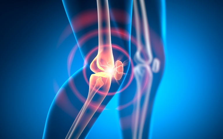
The role of extra-articular structures – particularly the collateral ligaments – in knee pain location and severity in osteoarthritis has been highlighted in a recent real-time ultrasound imaging study.
Knee pain in osteoarthritis is multifactorial and often poorly explained by structural imaging alone.
To address this, a recent cross-sectional study published in the journal Clinical Rheumatology examined associations among real-time ultrasound-detected structural pathologies, site-specific pain distribution and overall osteoarthritis pain severity.
The cohort comprised 54 patients (26 men, 28 women) aged 50 years or over with radiographic knee osteoarthritis and moderate-to-severe pain. Mean body mass index (BMI) was 31.5 kg/m²; 94–96% had Kellgren–Lawrence grade 2–3 disease, and average pain severity was 5.4 on a 0–10 numeric rating scale (NRS).
Participants underwent a detailed ultrasound assessment of extra-articular structures across all quadrants, including tendons, ligaments, bursae and Baker’s cysts.
Sono-palpation, which combines probe pressure with real-time imaging, was used to assess local tenderness. Pain location was mapped using the Knee Pain Map, aligning with ultrasound regions.
Ligament tenderness and osteoarthritis knee pain
The primary outcomes were the associations between ultrasound-detected pathologies and pain location and severity.
Medial knee pain was reported in 36 of 54 patients, consistent with involvement of the medial compartment in osteoarthritis. The most frequent ultrasound finding overall was hypoechogenicity of the semimembranosus tendon (53%), suggesting tendinopathy, although this was not associated with pain location.
In contrast, sono-palpation showed clearer anatomical correlations. Tenderness of the medial collateral ligament (MCL) was more common in patients with medial pain (43.2%), while lateral collateral ligament tenderness was more frequent in those with lateral pain (52.9%).
These findings were also linked to pain severity. MCL tenderness on sono-palpation was associated with a 1.41-point increase in the NRS score (95% CI 0.47–2.36; P=0.004), and Baker’s cyst tenderness with a 1.78-point increase (95% CI 0.35–3.20; P=0.016). A non-significant trend was observed for patellar tendon pathology (increase of 1.1 NRS points; P=0.08).
Potential clinical implications
The findings suggest that extra-articular structures may contribute meaningfully to osteoarthritis pain, both in terms of localisation and severity.
In addition, sono-palpation offers a novel method for linking imaging findings to clinically relevant tenderness, potentially improving diagnostic precision, the authors said.
They also proposed that ligament-related inflammation could reflect biomechanical stress around the knee joint and may be a therapeutic target – for example, through strategies aimed at mechanical unloading – but noted that some caution is needed when interpreting their results.
Pain is subjective and influenced by psychosocial and biological factors, which were not fully accounted for, thereby precluding causal inference.
Furthermore, the sample size was small, ultrasound assessment was performed by a single experienced operator, and intra-rater reliability was not evaluated, placing further limitations on the results.
The authors recommend that future research include larger, longitudinal studies incorporating both intra- and extra-articular assessments and evaluate biomechanical factors such as gait and alignment.
Including broader patient populations, such as those with structural osteoarthritis but minimal pain, will be important for confirming these findings, they added.
Reference
Ghouri A et al. The relationship between real-time ultrasound-detected extra-articular soft tissue pathologies and knee pain in osteoarthritis: a cross-sectional study. Clin Rheumatol 2026;45:2837–44.