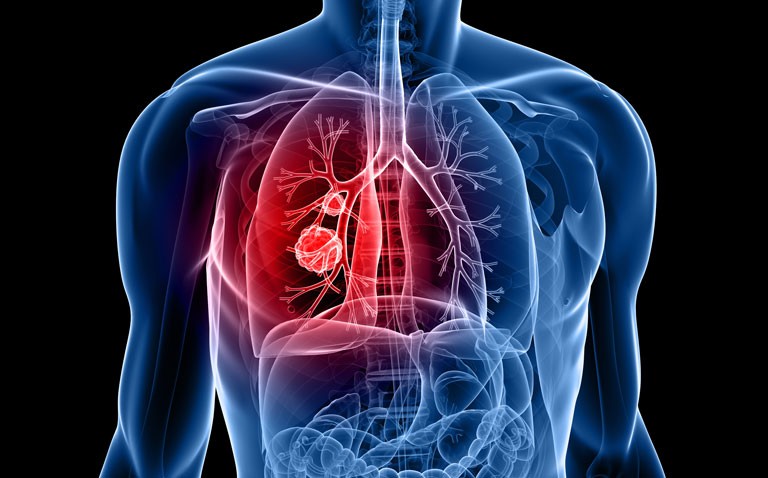
Combining infrared imaging with an AI system can determine if tissue samples contain cancer cells and identify lung adenocarcinoma.
Cancer is a leading cause of global deaths with lung cancer responsible for 11.4% of all cases and 18% of deaths. However, lung cancer is not a single entity and includes several subtypes such small-cell lung carcinoma, adenocarcinoma and squamous cell carcinoma, although adenocarcinoma is common, accounting for around half of all cases. In cases of suspected lung cancer, in particular adenocarcinoma, oncogene mutations are seen for at least 3 genes encoding for the proteins TP53, KRAS proto-oncogene and epidermal growth factor receptor (EGFR) and the presence of one of these mutations can influence the patient’s prognosis. The combination of laser-based Infrared microscopic imaging and microarrays has been used to examine colorectal tissue and to discriminate between malignant and non-malignant breast samples. This technique has been employed by a team from the Centre for Protein Diagnostics, Ruhr University, Germany, to determine the type of tumour present and mutations in lung samples. Initially, the team sought to train the AI system with samples of normal tissue and those from patients already diagnosed with adenocarcinoma. Once the system was able to correctly distinguish between normal and cancer tissue, a verification set was used with a much larger number of samples.
Data acquisition was performed with the laser infrared microscope on unstained tissue samples. Each pixel of the image was represented by one infrared spectrum and served as a morphological and molecular fingerprint for changes in the tissue. The AI system then classifies these spectra into different tissue types or molecular conditions and outputs the data as index colour images, where each colour represents a different tissue type or molecular condition. Pathological diagnosis is then made through histological methods such as immunohistochemical staining and next-generation sequencing with a gene panel to determine the mutation status.
Findings
A total of lung tissue sections from 235 patients with an average age of 68 years (101 female) were used in the verification set and of whom, 180 had been diagnosed with adenocarcinoma, 28 with squamous cell carcinoma and the remainder with other lung tumours. With respect to the determination of tissue type, the system had a sensitivity and specificity of 94% and 97% for adenocarcinoma. Similarly for squamous cell carcinoma, the system sensitivity and specificity were 96% and 96% respectively. Interestingly, for the other tumour types, which included carcinoid, neuroendocrine carcinoma and small-cell lung cancer, the sensitivity and specificity were all 100%. However, as the authors note, because there were a small number of tissue samples with these other cancers, the reliability of the results require further study. When examining the mutation status, the system correctly assigned more than 65% of the tumour spectra to the correct mutation class (i.e., KRAS, TP53 or EGFR).
In discussing their findings, the authors noted that while the system had a high degree of sensitivity and specificity for tumour type and mutation status, a further advantage was that the whole process could be performed in less than 30 minutes. They concluded that future work required a large number of patients to increase the reliability of the system and the augment the acceptability of the method in the medical community.
Citation
Goertzen N et al. Quantum Cascade Laser-Based Infrared imaging as a Label-Free and Automated Approach to Determine Mutations in Lung Adenocarcinoma. Am J Clin Pathol 2021;191(7):1269–80.


