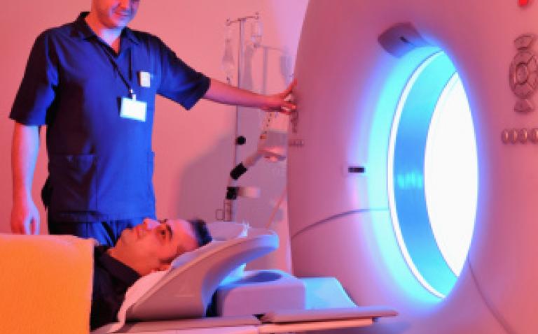
A unique collaboration between the Paul Scherrer Institute, CERN’s ISOLDE facility, and the Institut Laue-Langevin, has published preclinical study results for a newly developed set of tumour-targeting radiopharmaceuticals.
The results, published in The Journal of Nuclear Medicine, are a significant success for this group of nuclear medicine specialists and radiochemists, demonstrating their potential to research and even manufacture a new generation of radioisotopes with excellent properties for the diagnosis and treatment of cancer.
Background
‘Receptor-targeted’ radiopharmaceuticals [2] consist of a radioactive isotope which is attached to a biological component that selectively delivers it to tumour cells. They are used in two ways.
Nuclear medical imaging – involves injecting a positron or gamma-ray-emitting radiopharmaceutical into the bloodstream as a marker. Once bound to the target (e.g. a tumour), the gamma-ray emissions of the radioisotope or the positron annihilation [5] allow diagnosis and possibly the determination of the severity of a variety of diseases, including many types of cancers.
Targeted radionuclide therapy – makes use of short-range particle-emitting radioisotopes with the ability to destroy tumour cells.
Traditional radioisotopes (such as iodine-131 and yttrium-90), employed in the first generation of radiopharmaceuticals do not offer ideal nuclear properties for all therapeutic applications. As a result more recent developments of radiopharmaceuticals make use of emerging radioisotopes with more favourable decay properties such as lutetium-177 which reduces both collateral damage to healthy tissue and the need to isolate the patient during treatment.
The ideal situation would be to select the most suitable radioisotopes at an early stage in drug development, allowing an overall optimization of the radiopharmaceutical. However, such innovative radioisotopes are often not commercially available and dedicated production methods are often lacking.
Recently a method for large-scale production of one such new generation radioisotope, terbium-161, has been developed by radiochemists from TU Munich and PSI Villigen working with samples irradiated at the ILL in Grenoble and FRM2. These were successful in supplying this isotope in the quality and quantity needed for clinical applications.
In this latest study terbium-161 has been complemented by three other terbium isotopes, produced by high energy proton induced reactions at ISOLDE-CERN, which together have the potential to both diagnose and treat cancer. Having so called ‘matched pairs’ of isotopes (based on the same chemical element) is particularly valuable and opens up the opportunity for personalised, patient specific treatment to increase efficiency and reduce side effects. [4b]
Terbium (Tb) is the only element in the periodic table offering not only a matched pair but four clinically interesting radioisotopes with complementary nuclear decay characteristics which between them could be used in the full range of procedures in nuclear medicine.[5] Thus, terbium can serve as the “Swiss Army knife of Nuclear Medicine”, for fundamental studies of new radiopharmaceuticals and for detailed comparisons of targeted therapy options.
The trials
In a new joint article, scientists from the PSI, ILL and CERN reported on the first comprehensive preclinical study of this new range of terbium radiopharmaceuticals. The radioisotopes were combined with a newly developed delivery agent called ‘cm09’ [3] and then administered to tumour bearing mice. For the imaging isotopes, the scientists applied two common diagnostic techniques to study its uptake by cancer cells. Positron emission tomography (PET) and single-photon emission computed tomography (SPECT) were applied to the mice 24 hours after the injection of terbium-152, terbium-155 and terbium-161 respectively. Terbium-161 and terbium-149 were investigated with regard to their therapeutic efficacy by comparing tumour growth and survival rates in the mice under treatment with an untreated control group.
Key findings
PET/CT and SPECT/CT studies using both diagnostic isotopes terbium-152 and terbium-155 and the gamma-emitting therapeutic isotope terbium-161 provided excellent tumour visualisation 24 hours after injection.
Both therapeutic isotopes provided a significant inhibition to tumour growth in mice, resulting in a marked delay in tumour growth or even complete remission. In particular therapy with terbium-161 resulted in complete remission in 80% of cases.
The performance of terbium-161 was particularly encouraging as previous work by PSI, TUM and the ILL, published in 2011, demonstrated that this isotope could be produced in the quantity and quality required for clinical routine application.
Quotes
Dr Ulli Köster: “The development of new radiopharmaceuticals provides the chance of combining at an early stage new delivery agents with radioisotopes of optimum decay properties. Research facilities like ISOLDE at CERN and Institut Laue-Langevin can accelerate the development of promising new therapies by providing such radioisotopes in high quality that are not yet commercially available.”
Dr Karl Johnston, physicist at ISOLDE-CERN said: “ISOLDE is a nuclear facility at CERN which offers the greatest choice of radioisotopes of any such facility worldwide. Although the development of these radioisotopes was originally intended for fundamental studies in nuclear physics, these isotopes can often be applied to studies in materials science, biophysics and – increasingly – nuclear medicine. The application of radioactive isotopes to areas such as medicine is an exciting field which can also benefit society.
“This work demonstrates impressively how facilities that are mainly devoted to fundamental research such as ILL and CERN can accelerate the development of promising new cancer therapies.”
Notes and references
- Radioisotope production methods – Terbium-161 was produced by irradiation of gadolinium-160 targets with neutrons at Institut Laue-Langevin and Paul Scherrer Institute, converting it to the short-lived gadolinium-161 which in turn decays to terbium-161. Terbium-149, terbium-152 and terbium-155 were produced by irradiation of tantalum targets with high energy protons, followed by an online isotope separation process at ISOLDE/CERN.
- How to target tumours? – Overexpression of tumour markers (e.g. antigens, peptide receptors) which can be selectively targeted with biological vectors (e.g. antibodies or antibody fragments, peptides, vitamins) is a frequent characteristic of cancer cells. Such vectors can be combined with a payload, for instance a radioisotope
- cm09 – targets folate receptors. The folate receptor is overexpressed in a variety of aggressively growing tumours including ovarian and other gynaecological cancers as well as certain breast, renal, lung, colorectal and brain cancers, whereas its distribution in normal tissues and organs is highly limited. Folate vitamins show a rapid uptake but also a rapid renal elimination from the body, therefore it does not reside sufficiently long in the body to reach all cancer cells. Hence a new folate conjugate (indicated as ‘cm09’) was designed where folic acid is combined with an albumin binding entity that prolongs the circulation time in the blood.
- What is so great about terbium? 4a Terbium (Tb) is the only element in Mendeleev’s table offering not only a matched pair but even four clinically interesting radioisotopes with complementary nuclear decay characteristics covering all nuclear medicine modalities: terbium-152 for PET, terbium-155 for SPECT, terbium-149 for a-particle therapy and terbium-161 for therapy with electrons (b- , conversion and Auger electrons). Thus, terbium can serve as the “Swiss Army knife of Nuclear Medicine”, for fundamental studies of new radiopharmaceuticals and for detailed comparisons of targeted therapy options. 4b So-called “matched pairs” of a diagnostic and a therapeutic isotope of the same chemical element are particularly valuable since their identical chemical properties assure identical in vivo behaviour, enabling a precise determination and optimization of the radiation dose given to the tumour prior and during treatment. This opens the way for “theranostics”, where patients are first given a diagnostic isotope, then, based on the measured patient-specific uptake of the radiopharmaceutical, the optimum therapy option is selected and applied. This type of personalized medicine assures best possible efficacy and minimum side effects since the therapy is tailored to the patient’s needs.
- Different types of radiation are emitted by radioisotopes and their used in nuclear medicine: a Gamma radiation (emitted by terbium-155 and terbium-161) has a long range and will mainly escape the patient’s body. It can be detected with gamma cameras or SPECT scanners so is useful for monitoring exactly where in the patient’s body the radioisotope has been delivered. b Positrons (beta-plus particles emitted by terbium-152) will annihilate close to the emission point, creating two gamma rays that can be detected with PET scanners outside the patient’s body for monitoring exactly where in the patient’s body the radioisotope has been delivered. c Beta-minus radiation (emitted by terbium-161) has a range of few mm to cm and can damage or destroy cells in this range. d Alpha particles (emitted by terbium-149) have a range of few ten micrometers, comparable to a cell’s diameter and are more efficient in destroying individual cancer cells than beta-minus radiation. e Auger electrons (emitted by terbium-161) have a range of few micrometers only, shorter than a cell’s diameter. Their damaging effect is confined to a single cell, or even part of it. To be most effective Auger electron emitters need to be coupled to ‘internalising’ bioconjugates that are selectively incorporated into cancer cells
- About PSI – the Paul Scherrer Institute is Switzerland’s largest research centre for natural and engineering sciences. The multi-disciplinary centre focuses on three key areas: structure of matter; energy and the environment; and human health.
- About ILL – the Institut Laue-Langevin (ILL) is an international research centre based in Grenoble, France. It has led the world in neutron-scattering science and technology for almost 40 years, since experiments began in 1972. ILL operates one of the most intense neutron sources in the world, feeding beams of neutrons to a suite of 40 high-performance instruments that are constantly upgraded. Each year 1,200 researchers from over 40 countries visit ILL to conduct research into condensed matter physics, chemistry, biology, nuclear physics, and materials science. The UK, along with France and Germany is an associate and major funder of ILL. There are a further 10 scientific member countries.
- About ISOLDE-CERN: CERN is the European Organization for Nuclear Research based in Geneva. It is famous for its fundamental discoveries in particle physics, but its accelerator complex delivers also high energy protons to the ISOLDE facility. This isotope separation on-line facility provides mass-separated beams of over 1000 different radioisotopes. Some of these are of particular interest for innovative applications in nuclear medicine.


