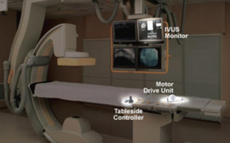
Stamford Hospital has employed the new iLab™ Ultrasound Imaging System to provide improved diagnosis and treatment of patients with coronary artery disease (CAD).
CAD, the leading cause of death for both women and men in the United States, affects more than 13 million Americans and is a result of atherosclerosis of the coronary arteries. Atherosclerosis occurs when arteries become clogged or narrowed by fatty deposits (plaque) that restrict blood flow to the heart. Without adequate blood, the heart is deprived of oxygen and the vital nutrients it needs to work properly.
The iLab Intravascular Ultrasound Imaging System uses high-frequency sound waves that reflect off tissue in the arterial walls and provide physicians with a cross-sectional view of the internal structure of the coronary arteries. Intravascular ultrasound shows physicians where the healthy arterial wall ends and the plaque begins. This three-dimensional image also helps doctors better understand the size and shape of a lesion blocking the artery, guides placement of stents (tiny mesh tubes used to restore blood flow to the heart) and provides information about how well the treatment worked.
“The iLab System provides an excellent view of the patient’s coronary artery disease from inside of the body, giving a clear understanding of the composition of the plaque and helping to determine the best diagnosis and treatment path,” said Dr Ted Portnay, medical director for Stamford Hospital’s cardiac catheterisation labs.
Intravascular ultrasound is performed during a cardiac catheterisation, when a catheter is inserted into an artery in a patient’s groin area and moved up to the heart. When this procedure is used in combination with angiography, the most common way to view arteries involving a two-dimensional X-ray, it provides a more complete picture of the diseased artery and helps the cardiologist to determine the best treatment option.


