

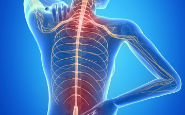


With increasing life expectancy and advances in cancer therapies, we face a population that presents a higher frequency of osteoporotic vertebral compression fractures (OVCF). The patient presents with pain due to vertebral fracture of a few weeks of development, which does not resolve with conservative treatment.
Percutaneous vertebroplasty (PVP) using polymethylmethacrylate was first performed by Deramond in 1984 and reported in literature in 1987.1 It is an image-guided procedure that involves the injection of acrylic cement into the bone. The aim is to strengthen the bone structure and to achieve pain relief and quality of life improvement (Figure 1).
The mechanism by which vertebroplasty reduces pain is still not completely known, but it has been shown that the bone cement produces greater strength, less distortion and reduced micromotion in the fractured vertebra. It also seems that there is a destruction of the sensory terminals by the cement matrix, as it is directly toxic by increasing the production of oxygen free radicals, and indirectly by the heat produced during cement polymerisation.
The ideal candidate for vertebroplasty is the patient who presents with a history of fracture of at least four weeks of development that occurs with back pain that increases with the burden of weight and is exacerbated by palpation of the spinous processes of the affected vertebra. However, clinical examination is not always so obvious. Many present with multiple fractures, which can hinder the determination of its age through routine diagnostic imaging techniques. Others present adjacent fractures, where clinical examination is difficult and does not allow determination of the cause of the symptoms.
Imaging techniques are useful in planning for vertebroplasty, regardless of whether the fractures are acute or chronic. Conventional radiology is useful but not definitive because many patients present with multiple vertebral compression deformities, so selecting the appropriate level to treat based solely on plain films can be problematic. Computed tomography provides greater anatomical detail, including or ruling out other structural elements at the source of pain (intervertebral disc, articular processes).
Magnetic resonance imaging (MRI) can detect the presence of bone marrow oedema (BME) in the fractured bone, such as increased intensity on T2-weighted images and hypointense signals on T1, reflecting the fluid inherent to the acute inflammatory response. Thus, as the fracture is resolved, the oedema disappears, and the signal is normalised on MRI, appearing isointense on T1 and T2-weighted images.2,3
The ability of MRI to identify BME has been studied in relation to the capacity of the technique in predicting the outcome of PVP in OVCF, acute and chronic. Brown et al.4 described the clinical benefits of PVP (improvement in pain and mobility) in 87% of the 45 patients with fractures over one year of development treaties, as 100% in patients with oedema MRI and found no direct correlation between MRI findings and the outcome of the PVP. Irani et al.5 presented the case of a patient with a fracture of five years of development, in which MRI showed an equivocal pattern showing no oedema (isointense on T1 with minimal increased signal in T2) and that PVP was effective.
Bone scans can provide additional functionality, as increased bone tracer uptake (uptake or overactive image) is shown in the images by the broken bone that increases its metabolic turnover in the attempt to heal. With this persistent bone remodelling, the damage by the fracture causes the pain.
Moreover, the new hybrid SPECT-CT equipment, combined scintigraphic images in tomographic acquisition (SPECT: single photon emission computed tomography) with radiological computed tomography (CT: computed tomography). This technology obtains functional and anatomical information together, and thus more accurately identifies the level of the symptomatic vertebra. Also, it provides greater anatomical detail and allows the assessment of other structural elements as the source of pain (intervertebral disc, articular processes). Collins et al.6 have described the benefits of combined SPECT-CT images compared to SPECT technology alone: additional information could be obtained in up to 62.5% of cases, resulting in significant changes in the final assessment.
The typical patient (Figure 2) presents with pain due to vertebral fracture after a few weeks of development, which does not respond to conservative treatment. MRI shows hyperintense short tau inversion recovery (STIR) T2-sequences translating bone oedema in the fractured vertebra. The bone scan shows increased tracer uptake in their images that increases its metabolic turnover in the attempted repair. The SPECT-CT fused images can find a higher resolution image on the crushed overactive vertebrae.
But it is not always good and different situations may hinder the correct diagnosis: the patient may present with a pain for a much longer time; a previous MRI may not demonstrate oedema; a recent fracture may produce a large structural instability or the MRI would not be performed for various reasons. In such instances, SPECT-CT can be useful.
In a previous study7 we retrospectively evaluated a series of 33 patients intended for PVP, in which treatment was highly efficient therapy for relief of pain due to vertebral fractures. We found that bone SPECT-CT would seem a good predictor of postprocedural response, that it would add valuable information to the cause of back pain, such as facet syndrome, and facilitates complete patient evaluation in patients that cannot receive MRI. However, formal conclusions about the prognostic value could not be drawn as there were insufficient SPECT-negative or pain-negative patients included.
In this scenario we proposed to carry out a new study to determine the value of bone SPECT-CT in response to PVP. It was a prospective, longitudinal, observational study that included 50 patients. Analysis included description and bivariate association between the predictive variable (bone SPECT-CT) and the outcome variable (clinical improvement). Collected variables were sex, age, number and level of fractures, and MRI scans using STIR sequences. A modified Oswestry Questionnaire (OQ), a visual analogue scale (VAS) for pain changes in analgesic treatment and clinical outcome, described baseline and follow-up data (at 3–12 months).
A total of 50 patients were included: 36 women and 14 men, aged 70.1 years (SD 8.4). On MRI, 47 patients presented with 83 vertebral levels displaying BME, while 96 vertebral levels presented increased uptake on SPECT-CT; 86.5% concordance. A total of 117 vertebral levels were treated (mean per patient 2.3; SD 1.6): 106 tested positive for either or both imaging methods, and 11 (negative for both) additional levels were prophylactically treated. Follow-up was obtained in 49 patients (mean 7.3; SD 4.5 months). A quantitative decrease in OQ pain score was noted: 30 to 17.5 points (p<0.001). Also a decrease was shown in the VAS pain score: 7.8 to 4.5 (p<0.001). Analgesic treatment was reduced from 2.7 to 1.2 (p<0.001) (0, don’t use; 1, self-medicated analgesics; 2, prescription of non-narcotic analgesics; 3, need for oral narcotics; 4, oral narcotics prescribed; 5, parenteral narcotics). Of the 50 patients treated, 26 had complete resolution of pain and 12 improved, while 10 patients unchanged and one patient worsened. Thus, pain relief was elicited in 38/49 patients (77.6%).
Of the 45 patients in whom a positive SPECT-CT was the basis to perform PVP, 38 had good analgesic response (84.4%), while seven patients had poor response (15.6%). On the other hand, in four patients whom SPECT/CT tested negative, a poor analgesic response was observed (100%). Therefore, SPECT-CT predicted pain relief in 85.7% (42/49) of patients treated by PVP.
From this study, our conclusions were that MRI and SPECT images showed high concordance (86.5%); pain relief was achieved in 77.6% of patients after PVP and that SPECT-CT predicted pain relief in 85.7% of patients treated.
In summary, two imaging techniques may be useful in the preoperative planning of PVP. MRI is currently considered the gold standard imaging technique in the preprocedural workup, based on its high sensitivity for BME and great anatomic detail. BME reflects the fluid inherent to acute inflammatory response. However, OVCF findings on MRI evolve over time, and chronic symptomatic fractures may show signal characteristics similar to those of normal bone marrow. SPECT-CT offers functional and anatomical information together and allows a better assessment of other structural elements as the source of pain, such as facet joints. Increased bone tracer uptake by the broken bone is due to increased metabolic turnover in an attempt to heal. Also, SPECT-CT can facilitate patient evaluation when MRI is not possible.
Each technique assesses a different aspect of the same process, which seems to concur in the same space and time. Both can help to select the candidate to PVP, as both give information about the functional and the anatomical situation of the broken bone. So, both may be useful to predict the outcome of a PVP and help to better define inclusion criteria of the procedure.

Figure 1.

Figure 2. Patient 1. A 68-year-old woman with four months spinal fracture. (left to right): Sagittal STIR MRI shows oedema in an acute compression fracture of L1; Planar scintigraphy presents classic horizontal radiotracer uptake; Sagittal CT image demonstrates L1 fracture; Sagittal and coronal SPECT-CT fused slices localises hyperactivity in the vertebral body L1.

Figure 3. Patient 2: MRI-STIR and SPECT fused sagittal images showing coincident oedema and hyperactivity in the L2 vertebral body.

Figure 4. Patient 3: A 81 year old woman: (left to right): Planar scintigraphy shows hyperactivity in L1 and L2; sagittal CT slice presents vertebral fracture of T11, T12 and L1; sagittal and axial SPECT-CT slices of L1 and L2 showing hyperactivity in the vertebral body of L1 and hyperactivity in L1–L2 facet arthroplasty. The patient reported pain relief after PV and injection of local anaesthetic.
References
- Galibert P et al. Preliminary note on treatment of vertebral angioma by percutaneous acrylic vertebroplasty. Neurochirurgie 1987;33(2):166–8.
- Modic MT et al. Degenerative disk disease: assessment of changes in vertebral body marrow with MR imaging. Radiology 1988;166:193–9.
- Do HM. Magnetic resonance imaging in the evaluation of patients for percutaneous vertebroplasty. Top Magn Reson Imaging 2000;11:235–44.
- Brown DB et al. Correlation between preprocedural MRI findings and clinical outcomes in the treatment of chronic symptomatic vertebral compression fractures with percutaneous vertebroplasty. Am J Roentgenol 2005;184:1951–5.
- Irani FG et al. Successful treatment of a chronic post-traumatic 5-year-old osteoporotic vertebral compression fracture by percutaneous vertebroplasty. Br J Radiol. 2005;78:261–64.
- Collins ZS, Nguyen MX, Bartley SC, et al. Comparison of SPECT/CT bone scans versus traditional SPECT bone scans in perioperative evaluation of vertebral compression fractures. Program and abstracts of the Radiological Society of North America 90th Scientific Assembly and Annual Meeting; November 28 –December 3, 2004; Chicago, Illinois. Page 699.
- Solá M et al. Value of bone spect-ct for chronic pain relief after percutaneous vertebroplasty in vertebral fractures. Spine J 2011;11:1102–7.
