


Pneumonia is a major cause of childhood mortality and morbidity globally – particularly in resource-poor environments – and diagnosis of pneumonia can be complex. The current gold standard diagnostic technique is a chest X-ray, looking for the presence or absence of areas of consolidation on the lungs.
However, this obviously exposes the patient to radiation, and X-ray facilities are not always available in remote locations. Dr Nathaniel Meuser-Herr led a University of Minnesota research project to investigate the potential of point-of-care ultrasound as an alternative tool for the diagnosis of pneumonia in paediatric patients in Uganda, and discussed the possibilities offered by this modality.
The University of Minnesota has a long history of paediatric research in Uganda, focusing primarily on malaria and malnutrition. As an offshoot of that work, we began looking into more practical aspects of medical care, such as how new or established medical technologies could be implemented as part of the local care network.
This resulted in a collaboration project with Hennepin Healthcare’s Emergency Ultrasound Fellowship and Global Health Uganda, funded by the Thrasher Research Fund, to evaluate the potential of point-of-care ultrasound as a diagnostic tool for ruling in or ruling out pneumonia in children presenting with clinical signs of the condition. This approach has already been trialled with some success in more controlled environments, so we wanted to see if it could be effectively implemented by a local healthcare team in Uganda.
Ordinarily, children presenting with clinical signs of pneumonia according to the WHO and Ugandan guidelines would be treated based on the clinical symptoms alone, and imaging would only be used if there were complications. We wanted to understand if the use of point-of-care ultrasound for every patient could improve both the overall diagnosis and patient care pathway.
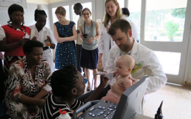
One of the key requirements for the study was having an ultrasound system suitable for use in a resource-poor environment, and so we approached FUJIFILM SonoSite. The company was kind enough to supply us with an M-Turbo system, which is ideally suited to this application, as it is compact and robust, and can run on batteries as and when required. It also has a relatively large screen, allowing several people to see the display at the same time for training purposes, which is vital for the sustainability of this type of programme.
Sonography training was provided to a doctor working in the Jinja Regional Referral Hospital, and all children presenting with signs of pneumonia for over a year – around 300 in total – were enrolled onto the trial. For each patient, both a chest X-ray and lung sonography were performed, and the programme also funded antibiotic treatments as appropriate.
The use of ultrasound was well received by patients and their parents, and one of the big advantages of ultrasound over X-ray in paediatric cases is that both the doctor and the parent can stay with the child during imaging, reducing the stress involved. The scans can also be done on the wards, without needing to transfer the patient to the radiology department, which further reduces anxiety.
We are currently in the process of analysing the data from this study, but point-of-care ultrasound certainly has the potential to help streamline paediatric pneumonia care in this type of setting – resource-poor hospitals that are already understaffed. Outside the scope of the study, the hospital’s medical team – once they had received the relevant training – were also able to take advantage of the availability of an ultrasound system for a range of other activities, using the M-Turbo to assess and triage inpatients and referrals.
They also used the system in a number of paediatric cardiology clinics, for both patient assessment and training purposes. We are very grateful to FUJIFILM SonoSite for supplying the M-Turbo system for use in this trial, as well as for subsequently donating it to Global Health Uganda. It is now providing vital imaging capabilities to a regional paediatric centre which has no access to other modalities, and will be available for future ultrasound research in the region.
