

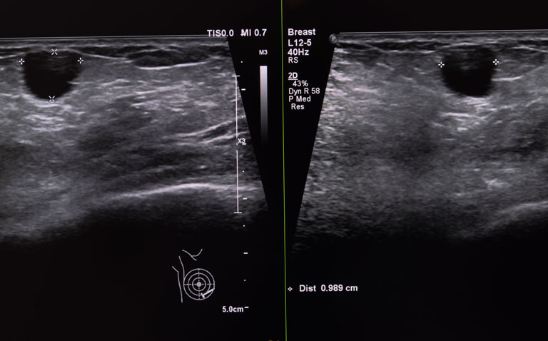


Diagnostic ultrasound is the imaging modality of choice for the assessment of intra-operative margin assessment of excised breast cancers
Diagnostic ultrasound appears to be the imaging of choice for an assessment of intra-operative margin assessment after excision of breast cancer according to a meta-analysis by researchers from Aarhus University Hospital, Denmark.
According to the World Health Organisation, in 2020 there were 2.3 million women diagnosed with breast cancer and globally, 685 000 deaths. The primary treatment modality for breast cancer is surgery with the aim of removing the cancer and conserving as much healthy tissue as possible.
However, there is currently a lack of consensus of an adequate margin in patients with invasive or in situ breast carcinoma undergoing breast-conserving surgery and over 20% of women require reexcision following their initial surgery.
Given the current reexcision rates, there is an obvious and important need for techniques to assess intra-operative breast margins. While frozen section and cytology have been shown to have the greatest diagnostic accuracy when assessing intra-operative breast margin assessment, such methods are resource intensive and turnaround times for results have prevented widespread adoption.
Although optical methods to determine margin status have been developed and with a high sensitivity and selectivity, no current modality has matured into a complete solution to the margin problem.
For the present study, the Danish team sought to evaluate the pooled diagnostic sensitivity, and specificity of radiological methods for intra-operative margin assessment and their impact on repeat surgery rate.
They performed a systematic search in PubMed, Embase, Cochrane Library, Scopus, and Web of Science for studies using several imaging techniques including radiography, digital breast tomosynthesis (DBT), micro-CT, and diagnostic ultrasound and where the histological assessment was used as the reference method.
The area under the curve (AUC) for each technique was calculated and a bivariate random effect model was used to obtained pooled sensitivity and specificity of the index tests in the meta-analysis.
Findings
The systematic search resulted in 22 articles with 29 radiological imaging methods and were included in the meta-analysis. Pooled sensitivity and specificity and area under the curve were calculated for a total of four subgroups: radiography, DBT, micro-CT and diagnostic ultrasound.
The AUC for radiography was 60%, DBT 76%, micro-CT 72% and diagnostic ultrasound 80%. The sensitivities were 52%, 67%, 68% and 72% for radiography, DBT, micro-CT and diagnostic ultrasound respectively. The corresponding specificities were 77%, 76%, 72% and 80%. However, the repeat surgery rate was poorly reported in the studies.
Although based on these findings, diagnostic ultrasound appeared to be the best imaging modality, the authors felt that use of ultrasound was not realistic for the current workflow processes and concluded that the ‘radiological methods for margin assessment need further improvement to provide reliable guidance in the clinical workflow and to prevent repeat surgeries.’
Citation
Manhoobi IP et al. Diagnostic accuracy of radiography, digital breast tomosynthesis, micro-CT and ultrasound for margin assessment during breast surgery: A systematic review and meta-analysis. Acad Radiol 2022.
