


Contrast-enhanced computed tomography (CT) assessment of liver enhancing tumour burden in those with multifocal neuroendocrine liver metastases, enables predictions of patient outcomes, according to a retrospective study by a team of European researchers.
Neuroendocrine tumours are a heterogenous group of cancers most often in the gastrointestinal or respiratory tract. Although neuroendocrine metastases are rare, hepatic metastases can occur in as many as 75% of patients, which significantly worsens their prognosis.
There are several intra-arterial therapeutic options available to treat metastatic disease and while the evidence base was deemed of moderate quality, these approaches have been endorsed in consensus guidelines.
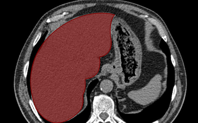
Volumetric assessment of metastatic disease in the whole of the liver, is known as the liver enhancing tumour burden and provides a means of evaluating a patient’s response to treatment following intra-arterial therapy.
However, in most cases, these assessment have been undertaken using MRI. In the resent study, researchers investigated the value of contrast-enhanced CT to assess liver enhancing tumour burden and to explore whether changes in this burden might serve as an early response marker to help predict survival outcomes for patients with multifocal neuroendocrine liver metastases after intra-arterial therapy.
The researchers retrospectively identified individuals with neuroendocrine liver metastases who had undergone intra-arterial treatment with either transarterial embolism or chemoembolism and who had contrast-enhanced CT scans. The liver enhancing tumour burden was quantified before treatment and then at the first post-treatment CT scan. The researchers calculated overall survival (OS) and progression-free survival (PFS) using Cox proportional hazard analysis.
Contrast-enhanced CT and treatment outcomes
A total of 119 patients with a mean age of 60 years (51.2% male) and who underwent 161 treatments were included in the analysis. Contrast-enhanced CT revealed a median pre-treatment liver enhancing tumour burden of 17%.
Following intra-arterial treatment, the liver enhancing tumour burden reduced by a median of 25.8%. This change was the best discriminator for overall survival (83 months among responders compared to 51 months in non-responders, p = 0.02) and for whole-body progression-free survival (18 months vs eight months, p < 0.001). The change in liver enhancing tumour burden was found to be independently associated with improved OS (hazard ratio, HR = 0.56, 95% CI 0.33 – 0.95, p = 0.03).
In addition, a 10% reduction in liver enhancing tumour burden best predicted the time to when intra-arterial treatment would be unfeasible (HR = 0.44, 95% CI 0.28 – 0.70, p = 0.01).
The authors concluded that the use of contrast-enhanced CT assessment of liver enhancing tumour burden, could help to predict survival outcomes in patients with neuroendocrine metastases following intra-arterial treatment.
They also proposed that future studies should focus on whether their approach might be generalisable to enable an assessment of other treatment modalities and systemic treatments.
Citation
Assouline J et al. Volumetric Enhancing Tumor Burden at CT to Predict Survival Outcomes in Patients with Neuroendocrine Liver Metastases after Intra-arterial Treatment. Radiol Imaging Cancer 2023.
