


Results from a randomised controlled trial involving 300 prostate cancer patients find that a molecular imaging technique is more accurate than conventional medical imaging.
The research recommends the scans be introduced into routine clinical practice.
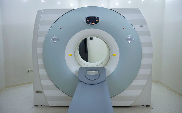
A medical imaging technique known as PSMA PET/CT that provides detailed body scans while detecting levels of a molecule associated with prostate cancer could help doctors better tailor treatments for their patients, by determining the extent of disease spread at the time of diagnosis, a randomised controlled trial involving 300 patients in Australia published in The Lancet journal has found.
The approach combines two imaging technologies – positron emission tomography (PET) and computed tomography (CT) – and is almost one third more accurate than standard imaging at pinpointing the spread of prostate cancer throughout the body. PSMA PET/CT proved to be 92% accurate compared with only 65% accuracy with standard imaging.
Although the study did not assess whether the scans had any effect on patient survival, the researchers say this approach could improve outcomes by helping doctors decide whether to offer a localised treatment, such as surgery or radiotherapy, or to use more advanced treatments to treat the whole body if the cancer has already spread.
Costs associated with PET/CT vary by geographical area and the researchers caution that a full economic analysis will be critical for determining the feasibility of widespread use. Nevertheless, they recommend a review of current clinical guidelines and for PSMA PET/CT to replace the use of conventional imaging where possible for men with high risk prostate cancer.
Prostate cancer is commonly treated by surgery to remove the prostate or intensive radiotherapy to target the tumour. If there is a high risk the cancer may have spread to other parts of the body, patients may be offered medical imaging – typically CT and bone scans – to help doctors determine if additional treatments are needed.
Study lead Professor Michael Hofman of the Peter MacCallum Cancer Centre, Australia, said: “Taken together, our findings indicate that PSMA-PET/CT scans offer greater accuracy than conventional imaging and can better inform treatment decisions. We recommend that clinical guidelines should be updated to include PSMA PET/CT as part of the diagnostic pathway for men with high risk prostate cancer.”
Researchers sought to investigate if a molecular imaging approach could help doctors better define the extent of disease at the time of diagnosis. This approach involves giving patients a radioactive substance that detects prostate specific membrane antigen (PSMA), which is found at high levels on prostate cancer cells. They then undergo a PET/CT scan. The CT scan produces detailed images of the body’s organs and structures, while the PET scan lights up areas where PSMA is present at high levels, indicating the presence of prostate cancer cells.
The study involved 300 men recruited to ten sites across Australia. All of the men had been diagnosed with prostate cancer, confirmed by tests on prostate tissue samples, and were deemed to be at high risk of having aggressive disease. The men were randomly assigned to receive either conventional CT and bone scans (152 patients) or PSMA-PET/CT (148 patients). Men then swapped over and were given the scans using the alternative imaging arm unless more than three sites of cancer spread were detected on the initial scans (18 patients). A low number of men dropped out of the study or follow up information was not available for them (five patients). The remainder were given a second round of medical imaging at their six month follow-up appointment. The results of these scans were used to confirm tumour spread, in addition to biopsies and change in blood tests.
Overall, the researchers found the PSMA-PET/CT scans were much more accurate than conventional CT and bone scans at detecting cancer spread (92% vs 65%). This is because the new technique was better at detecting small sites of tumour spread. Conventional imaging failed to detect that the cancer had spread in 29 patients, giving a false negative result. By comparison, PSMA-PET/CT gave false negative results in just six patients. Furthermore, fewer men had false positive results obtained with the new technique (2 with PSMA-PET/CT and 9 with conventional imaging).
Patients who underwent PSMA-PET/CT scans had fewer ambiguous results than conventional imaging (7%, 11/148 patients vs 23%, 35/152 patients).
Both imaging techniques involve exposure to radiation but the dose associated with PSMA-PET/CT was less than half that associated with conventional imaging (8.4mSv vs 19.2mSv).
PSMA-PET/CT scans had greater impact on the way patients’ disease was managed, with 28% having their treatment plans changed after the scans (41/147) compared with 15% following conventional imaging (23/152).
When PSMA-PET/CT was given at the second round of imaging after conventional imaging, disease management plans were still changed in more than a quarter of cases (39/146, 27%). When conventional imaging was used at the second round, however, just 5% of patients had their treatment plans changed (7/135 patients).
Professor Declan Murphy, senior author, said, “Around one in three prostate cancer patients will experience a disease relapse after surgery or radiotherapy. This is partly because current medical imaging techniques often fail to detect when the cancer has spread, which means some men are not given the additional treatments they need. Our findings suggest PSMA-PET/CT could help identify these men sooner, so they get the most appropriate care.”
Associate Professor Roslyn Francis, co-author, of the University of Western Australia, said: “Costs associated with PSMA-PET/CT vary in different regions of the world but this approach may offer savings over conventional imaging techniques. A full health-economic analysis will help to determine the cost effectiveness of introducing PSMA-PET/CT, both from a patient and a healthcare perspective“
Some patients in the study had undergone further tests to confirm the spread of disease, which involved removing tissue from their pelvic lymph nodes. This is acknowledged as the most reliable test for assessing the stage of prostate cancer disease spread. The researchers caution that as not all patients underwent the procedure, it may lead to an overestimation in the sensitivity of PSMA-PET/CT scans at detecting smaller tumours. However, as patients had been randomly assigned to their groups, the researchers conclude that their study provides robust comparative data.
Writing in a linked Comment article, Professor Caroline Moore, University College London (who was not involved in the study), said: “Introduction of novel imaging into routine practice requires careful assessment of the potential burden to both individuals and to society, taking into account changes in treatment which can result. The proPSMA authors have planned an economic analysis of the potential effect of replacing conventional staging with PSMA-PET, which will be crucial in assessing the feasibility of widespread use of PSMA PET-CT in men being considered for radical treatment for high-risk prostate cancer.”
