Regular endoscopic surveillance of Barrett’s oesophagus has long been the standard of care, but it is costly, invasive and burdensome for patients. Here, Dr W. Keith Tan describes how a simple capsule-sponge test validated in real-world settings offers a breakthrough in precision risk stratification, paving the way for more efficient and patient-friendly surveillance of those most at risk of progression to oesophageal cancer.
Barrett’s oesophagus is the only recognised precursor to oesophageal adenocarcinoma – a cancer whose incidence has risen sharply over the past three decades. Although progression from Barrett’s oesophagus to cancer is relatively uncommon, when it does occur, the consequences are serious. The key aim is therefore to detect dysplasia or early stage cancer at a stage when endoscopic treatment can still be curative.
If oesophageal cancer develops, the treatment is an oesophagectomy, where a surgery is performed to remove the oesophagus and part of the stomach. This surgery is, however, a high-risk procedure and associated with significant morbidity and mortality.
Regular endoscopic surveillance of Barrett’s oesophagus is therefore a safety net to detect early cancerous changes and to prompt timely intervention, but it is blunt and resource-intensive if everyone is treated the same.
The holy grail of Barrett’s oesophagus surveillance is accurate risk stratification, which would enable the identification of those who are more likely to progress to cancer. Currently, however, all Barrett’s oesophagus is treated the same. Hence, a well-tested and validated risk stratification strategy would enable the transition from a one-size-fits-all model to precision surveillance. Here, high-risk patients would be fast-tracked for diagnostic endoscopy and treatment, while low-risk patients could be monitored less invasively, thereby reducing unnecessary procedures, anxiety and costs.
Limitations of endoscopic surveillance in clinical practice
Endoscopy is a resource-intensive process that requires specially trained operators. The procedure also often requires sedation, and the patient may therefore need to take time off work. It also utilises pathology capacity, which is both time-consuming and costly.
Endoscopic quality is also variable in real-world practice, where adherence to systematic biopsy protocols is inconsistent, and only around 50% of people adhere to the gold standard surveillance practice.
For the majority of Barrett’s oesophagus patients, serial endoscopies represent a substantial burden economically, without proportional clinical yield, given that only a small proportion of cases progress to cancer. Waiting lists and capacity constraints exacerbate this issue, potentially delaying patients who require urgent attention.
Furthermore, endoscopy is not without risk. There is a risk from the sedation itself, as well as a risk of bleeding and perforation of the viscus, which can sometimes be life-threatening.
A promising strategy for routine monitoring
The capsule-sponge test is a simple, quick and scalable technique that can be easily performed in an outpatient or primary care setting.
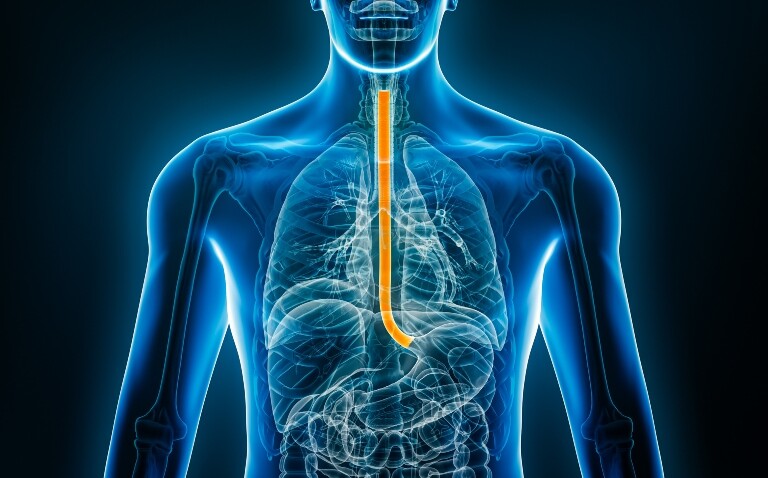
The patient swallows a pill-sized capsule, and once it enters the stomach, the vegetarian gelatine vehicle disintegrates to release a small mesh sponge 3cm2 in diameter, which, when withdrawn, can retrieve approximately one million cells from the oesophagus.
The test is simple to administer, and limited training is required to achieve competency. It can be delivered in an outpatient setting in under 10 minutes, without sedation or recovery time.
The sponge samples can then be stained with biomarkers that can be interpreted objectively (i.e., yes or no), including Trefoil factor 3 for goblet cells, assessment of cytological atypia, and p53 immunohistochemistry, enabling reproducible risk stratification while minimising operator dependence and costs.
A landmark study
Previous studies of the capsule sponge had shown promise in controlled, research-focused cohorts. However, healthcare systems require solutions that function effectively in everyday clinical practice, where there is heterogeneity among operators, pathologists and patient populations.
Our recent study, published in The Lancet, was conducted across routine NHS clinics, with real-world workflows, and yet the classifier still performed robustly. This gives confidence that the approach is not just an academic exercise but something that can be scaled and implemented within standard care pathways.
The classifier model itself is deliberately simple: it integrates three clinical variables, such as age, sex, and Barrett’s segment length, with biomarker readouts for cytological atypia and p53 status from the capsule sponge.
Despite its simplicity, it was able to meaningfully separate patients into low-, moderate- and high-risk groups. Crucially, it identified most patients with prevalent dysplasia or early cancer, who could then be prioritised for immediate endoscopy and potential curative treatment.
A paradigm shift in Barrett’s oesophagus surveillance
The real significance of our study is that it demonstrated, for the first time in a prospective and pragmatic setting, that a non-endoscopic test could reliably stratify patients with Barrett’s oesophagus according to their risk of harbouring or progressing to dysplasia and early cancer.
This represents a significant advancement in surveillance. Up until now, every patient with Barrett’s oesophagus has essentially been treated the same, scheduled for endoscopy at fixed intervals – either three- or five-yearly – regardless of their actual risk.
This blanket approach is costly, inefficient and in some cases, unsafe, as it can delay endoscopy for patients who need it most. By embedding the capsule-sponge test at the front of the pathway, we can invert this model: low-risk patients are safely monitored with a simple outpatient capsule-sponge test, while high-risk patients are escalated rapidly to diagnostic endoscopy.
In short, the significance lies in two key aspects: it provides real-world validation, proving feasibility at scale, and empowers us to move towards a precision surveillance model in Barrett’s oesophagus that is more efficient, patient-friendly and ultimately more effective at catching early cancers when they are still curable.
Allocating endoscopy resources more effectively
This is one of the most impactful findings from the study. Our data show that over half of the surveillance population could be confidently classified as low risk using the capsule-sponge test. This is not a trivial proportion; it represents thousands of patients each year who could safely avoid repeated endoscopies.
The implications are threefold. First, it frees up capacity in overstretched endoscopy units. For example, the waiting list for an upper endoscopy in most UK hospitals is three to six months. By reducing routine procedures in low-risk patients, we create space for urgent diagnostic endoscopies, therapeutic interventions and higher-risk surveillance cases.
Second, it reduces the burden on patients. Endoscopy requires preparation, sedation in many cases, and time off work. Avoiding repeated invasive procedures spares patients the anxiety, inconvenience and potential complications associated with lifelong surveillance. Instead, they can be followed with a simple 10-minute outpatient capsule-sponge test, which provides reassurance without disrupting their daily lives.
Third, it improves value for health systems. Endoscopy is costly both directly and indirectly, whereas the capsule-sponge is inexpensive, rapid and nurse-led. Redirecting resources away from unnecessary procedures enables healthcare systems to deliver more care for the same expenditure while maintaining safety.
Importantly, ‘low risk’ does not mean ‘no follow-up’. These patients are still monitored with the capsule sponge, and if they are found to have high-risk test results, then endoscopy is warranted. It represents a more rational, precision-based allocation of healthcare resources, ensuring the right test is given to the right patient at the right time.
The role of multidisciplinary teamwork
Multidisciplinary collaboration is absolutely central to making this model work at scale. The capsule-sponge test itself may be simple but embedding it into Barrett’s oesophagus surveillance pathways requires coordinated input across the entire care team.
Nurses are at the frontline. They can be trained quickly to deliver the test safely and consistently in outpatient clinics, ensuring accessibility and standardisation.
Pathologists play a crucial role in interpreting the samples, particularly in reporting atypia and p53 status with reproducibility. We, however, plan to move towards artificial intelligence (AI) interpretation of the sponge samples, and work is underway to automate this process.
Gastroenterologists and endoscopists remain vital, but their role shifts. Instead of performing surveillance on all patients, they can focus their expertise on those flagged as high risk, performing diagnostic and therapeutic endoscopy where it matters most.
Surgeons, oncologists and radiologists are then engaged earlier in cases where dysplasia or cancer is detected, ensuring patients move rapidly into treatment pathways.
Enhancing surveillance strategies for Barrett’s oesophagus
There are three main areas of focus. The first is biomarker refinement, utilising multiomic panels and quantitative scoring to refine risk prediction and interval safety. The holy grail of Barrett’s oesophagus risk stratification is to diagnose the condition and, at the time of diagnosis, predict whose disease will progress and whose disease will remain dormant.
The second area is AI and digital pathology employing assisted grading of atypia and p53, alongside automated reports to reduce variability and turnaround times.
The third area is trials to prove that surveillance is beneficial. Currently, although most international guidelines recommend surveillance, the effectiveness of this approach is uncertain, and there is an increasing recognition that the current surveillance strategy is ineffective.
Nonetheless, while guidelines still recommend surveillance, the capsule sponge offers a far better, more patient-friendly and resource-efficient alternative to endoscopy.
The future of Barrett’s oesophagus
We see potential for this model being adapted for monitoring other pre-cancerous conditions or applied to different tumour types. The broader principle is that less invasive tests, coupled with robust biomarkers, can replace repeated endoscopy or imaging.
We have shown this in Barrett’s oesophagus with the capsule sponge, but similar approaches could be developed elsewhere. For example, blood-based biomarkers or volatile organic compounds in exhaled breath are being investigated as non-invasive tools for identifying cancers of the lung, stomach and oesophagus.
The key goal is to shift from invasive, resource-heavy procedures towards patient-friendly tests that still deliver accurate risk stratification and early cancer detection.
Author
Dr W. Keith Tan Bsc (hons) MBChB MRCP PhD
Honorary registrar in gastroenterology and hepatology, Addenbrooke’s Hospital, and clinical researcher at the Early Cancer Institute, University of Cambridge, UK