Dr Matthew Webber, a clinical training fellow at University College London’s Institute of Cardiovascular Science, is co-developer of a reusable heart vest that can map the electrical activity of the heart in fine detail. He speaks to Katherine Price about how this innovation could potentially be used to better identify people at high risk of sudden cardiac death.
Around two million people experience heart rhythm disorders in the UK, according to the NHS, and 500 people die every year from sudden cardiac arrest.
‘Often, there was no particular warning,’ says Dr Matthew Webber, who works at Barts Heart Centre in London – a centre of excellence for cardiovascular care and the largest cardiac centre in the UK. ‘Usually, these patients will either have genetic rhythm problems, which they’re born with, or heart muscle disorders that they’re either born with or develop.’
Genetic heart rhythm disorders and cardiomyopathy can be difficult to detect, and it is similarly hard to predict risk of sudden cardiac death, as it is not known how structural features or abnormalities of a heart can influence risk.
Although medication or implantable cardioverter-defibrillators can prevent sudden cardiac events, it is challenging for cardiologists to assess who is at risk and who is not.
Cardiac magnetic resonance (CMR) imaging can show the health of the heart tissue, but not its electrical activity, and so Dr Webber says that ‘to complete that picture, you need to overlay the electrical information onto the heart scan to help better establish that risk’.
A 12-lead electrocardiogram (ECG) can help, as it’s a quick and cheap procedure, but it hasn’t really advanced over the last 50 years. It also involves applying electrodes with gel, which can irritate the skin and be uncomfortable, and the image resolution also isn’t high enough to provide more detail than the general location of where in the heart a problem is coming from.
Electrocardiographic imaging innovation
Until recently, detailed mapping of the heart’s electrical activity was rare, requiring either a catheter to be inserted inside the heart cavity or the use of expensive, single-use devices that were time consuming to set up and involved radiation.
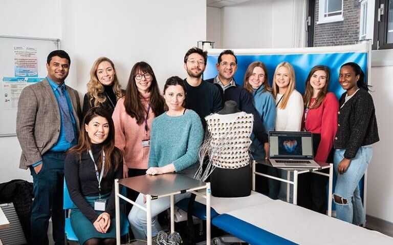
But that has all changed thanks to some innovative thinking from Dr Webber and colleagues at the UCL Institute of Cardiovascular Science, who are pictured above.
It was Dr Gaby Captur (pictured sitting, centre), consultant cardiologist in inherited heart muscle conditions at the Royal Free London NHS Foundation Trust and senior lecturer at University College London’s (UCL) Institute of Cardiovascular Science, who had the initial idea: developing an electrocardiographic imaging (ECGI) vest that could be used alongside MRI, and she brought Dr Webber on board as co-developer.
‘We needed to invent a vest that could be used with cardiac MRI, was reusable, cheap, quick, safe and effective, could be used on large numbers of patients for research, and then hopefully for clinical use as well,’ explains Dr Webber. ‘We could [then] complete this picture of heart disease by incorporating the electrical map of the heart with the cardiac MRI.’
The team received a grant from the British Heart Foundation (BHF) and the Society for Cardiovascular Magnetic Resonance to develop the concept, working with UCL BHF clinical research training fellows Dr George Joy and Dr Fiona Chan.
Designing and testing the heart vest
The design of the vest was based on the layout of the only commercially available ECGI vest - Medtronic’s CardioInsight single-use vest, which is compatible with computed tomography.
Conventional single-use ECG electrodes are made from silver-silver chloride and used with electrolyte gel. For the vest, the team developed bespoke dry electrodes from silver-coated polyamide yarn and the vest is fitted with 256 of these on the front and back.
The team tested the electrodes in-house for electrical characterisation, ECG signal quality, surface resistivity and washability. The textile-based sensors use conductive polymers that don’t require gel and can be washed and therefore re-used – the first time such electrodes have been used for ECGI.
The vest itself is made from cotton for its durability, comfort and ECG signal quality and was designed to fit a range of body habitus and size. It was also tested for its suitability for the CMR environment and to be machine washable up to 100 cycles.
The recording process takes approximately five minutes, requiring around 15 minutes of processing time in total per patient.
‘Anyone can do the recording, it doesn’t really require any special skill, just a bit of training on the software, and then patients go and have their cardiac MRI scan after that,’ says Dr Webber. ‘This can be done in one sitting in one centre and it doesn’t rely on the patient moving around or having an invasive test to complete that cardiac examination.’
Initially, Dr Webber had to manually combine the electrical data from the vest’s sensors with detailed images of the heart structure taken by MRI to generate 3D digital models of the heart and its electrical activity. The Barcelona Supercomputing Center then came on board to support the development of more efficient semi-automated techniques.
The last piece of the puzzle
Before 2020, studies mapping the electrical activity of the heart had 20 or 30 patients at most. But, once up and running, Dr Webber and the team assessed the vest’s feasibility in 77 patients.
The results, published in the Journal of Cardiovascular Magnetic Resonance, found it to be reliable and durable, with good reproducibility in younger and older participants.
The vest has since been used on 800 patients, and a larger study mapping the hearts of people with diseases such as hypertrophic cardiomyopathy and dilated cardiomyopathy is due to be published in the journal Circulation.
‘We had pretty much 100% tolerability in terms of safety and comfort. Some patients couldn’t necessarily lie flat for five minutes if they had back problems, but that was fairly limited,’ says Dr Webber. ‘The patients commented that they felt it was comfortable and easy and quick.’
He adds: ‘Out of the 800 cases we did, we had around 10 or 15 that couldn’t be analysed, so a very small number that dropped out of the analysis for various reasons.’
By making high-throughput, non-invasive and radiation-free CMR-ECGI possible, the vest could open the door to making whole-heart panoramic electrophysiological mapping suitable for healthcare and large-scale population research studies.
It may also provide insights into arrythmia, potentially paving the way for more personalised risk stratification in patients with heart muscle disease.
‘We hope that the vest can be a cost-effective screening tool, and the rich electrical information that it provides can help us better understand patients’ risk of heart rhythm disorders and help with treatment of those,’ says Dr Webber. ‘It can also be used to assess the impact of drugs and lifestyle interventions on the heart. It completes that overall picture, it’s that missing link.’
The future of the heart vest and sudden cardiac death
It’s early days, and potential biomarkers obtained through CMR-ECGI that could predict risk of sudden cardiac death and heart rhythm disorders will need to be monitored over time to assess the relationship to outcomes.
For now, the vest has been patented and the FDA approval process is underway. The team hopes to sell it to research groups before rolling it out clinically within the next five years.
As wearable health technology and artificial intelligence become more prevalent and change the way results are taken and risk is identified, the vest’s potential is seemingly infinite.
‘Using smart wearable devices is going to be a big deal, allowing people to wear diagnostic devices at home like smart watches so they don’t have to go to hospital,’ concludes Dr Webber.
‘Smart wearable devices and using automated AI to help establish patients’ risk is going to be the way forward.’
Image courtesy of UCL Institute of Cardiovascular Science / James Tye.