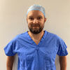

Vince Walker has worked at the Royal Stoke Hospital for 14 years and is currently head of cardiac arrhythmia diagnostics and electrophysiology.
Training as a cardiac physiologist in 2009, Vince Walker has a particular interest in technology in heart care and has since completed a postgraduate accreditation with the British Heart Rhythm Society (BHRS) and the International Board of Heart Rhythm Examiners (IBHRE) for both cardiac devices and cardiac electrophysiology.
Walker is passionate about using technology within the NHS to help manage patients with detectable conditions, such as atrial fibrillation (AF), to deliver “cost-effective, accurate and swift diagnostics to patients”.
He shares how he helped evolve the stroke care pathway at his hospital and why tech and diagnostics are the future of healthcare.
Why is managing patients with detectable conditions, such as AF, so important?
The link between AF and stroke is established but not fully understood. The immediate and long-term effects of a stroke on an individual, their family and the NHS is significant. It is fiscally costly to treat and manage a patient following an embolic stroke and, more importantly, the personal cost to an individual patient is often devastating. However, there is a general acceptance that AF-related stroke is largely preventable with prolonged cardiac monitoring to detect AF and anticoagulation therapy.
Looking at technology in heart care, you helped evolve the stroke care pathway at your hospital. Please can you tell us more about it?
I worked closely with Dr Indira Natarajan and Rachel Powell in neurology stroke services to develop a pathway for post-TIA/stroke patients to access implantable cardiac monitors as an option to help to detect AF after cryptogenic stroke. In the wider stroke population, conventional arrhythmia diagnostics using ambulatory monitoring fails to reveal a high incidence of AF and cannot rule out paroxysmal AF as a risk factor for stroke. This equates to approximately 25% of referrals into the arrhythmia monitoring service.
A small pilot study at the Royal Stoke Hospital comparing 50 post-stroke patients inserted with the device to a post-stroke cohort fitted with conventionally ambulatory monitors (observed retrospectively over six months) was found to have considerably different diagnostic yields to detect AF (15% vs <1%). This led to the development of our direct access service and the use of these insertable heart monitors in the most appropriate post-stroke patients.
What advantages did this bring?
It led to expedited diagnosis of AF and intervention with anticoagulation compared to traditional follow-up methods. Waiting times are often shorter for device implantation compared to ambulatory monitoring.
The pathway extends to patients that have made a good recovery post-stroke and would be considered for anticoagulation if AF was detected. There is also the additional therapeutic value in patients diagnosed with AF; it may be the case that symptoms related to the rate of arrhythmia occur as events become more frequent. We are currently observing patients with detected AF and their long-term outcomes following treatment with respect to drug therapy, ablation or even pacemaker therapy.
How were patients managed before the introduction of this pathway?
Post-stroke patients would be referred for 24-hour ambulatory or Holter cardiac monitoring. This would be performed approximately six weeks later in an outpatient clinic. The results of the monitors would generally be reported back to the stroke team within seven days. If AF was not detected it would be typical to offer longer-term monitoring of up to seven days, but this would often require a further waiting time of six weeks or longer. Essentially, referrals for prolonged cardiac monitoring may typically require several months of waiting and still be very unlikely to diagnose AF.
Has this approach led to cost savings?
The cost savings are difficult to appreciate through secondary care budgets. The stroke physicians believe the biggest cost saving is through the reduction of AF-related stroke by means of anticoagulation, better patient outcomes and lesser stroke severity, which, if reduced, may show more of a cost saving in the social care sectors. In a related study, the approach led to a 64% reduction in risk of stroke and a 25% reduction in mortality. Regrettably, this pathway is geared toward patients who have already transited secondary care treatment for which the initial cost has already been incurred. The evidence for device-detected AF, anticoagulation and relative stroke risk reduction is currently unclear, but, by developing pathways, we hope trends will emerge to further these pathways in the future.
How did you develop your expertise in this area?
I spent two years [2015-2017] working to consolidate the more technical aspects of cardiac devices and to really understand the potential and limitations of device care with patients. I now lead a large cardiac rhythm management (CRM) diagnostics service at University Hospitals of North Midlands NHS Trust (UHNM) that delivers approximately 1,200 conventional ambulatory ECG monitors and 50 insertable devices per month through a team of 15 specialist arrhythmia physiologists and clinical scientists.
What does a typical day look like for you?
Typically, I work an 8am to 5pm day with a one-in-seven on-call out-of-hours and overnight. I hold a clinical role within cardiac devices and electrophysiology covering outpatient clinics and procedures undertaken in the cardiac catheterisation lab. In addition, I am the service lead for electrophysiology and cardiac rhythm management and lead a large team to deliver a nationally recognised service. I would not be in this position without such a motivated, enthusiastic and hardworking team aligned to the same goal.
How is your hospital set up for cardiology medicine?
Roles range from Band 2 technicians with a primary focus on the delivery of ambulatory monitors and 12-lead ECG in outpatient settings and on the front line in A&E to coordinate flow through other outpatient clinics such as echocardiography. Band 3 and 4 staff complete a similar role but with more responsibility and independence, often working out in the community where specialist support is not readily available.
Then Band 5 cardiac physiologists provide the majority of the more complicated tests, such as exercise tolerance tests, and are involved in cardiac catheterisation labs and the reporting of ECG data. Band 6 roles take on further responsibility within arrhythmia monitoring and are considered entry-level positions in postgraduate training opportunities within echocardiography or cardiac devices.
Finally, Band 7 members of staff are qualified within those advanced practice roles to provide the more complex tests and reports to diagnose or contribute to the diagnosis of cardiac conditions.
What procedures or treatments are carried out most frequently?
On a weekly basis, UHNM NHS Trust’s cardiac diagnostics department provides more than 500 echocardiograms, over 300 ambulatory monitors, more than 300 ECGs, 500 insertable transmissions and in excess of 500 pacemaker/defibrillator appointments.
The wider team comprises nearly 70 staff delivering 2,000 patient interactions per week. Organising this many investigations is challenging given the flexibility of the NHS, the time sensitivity required for certain inpatient tests and the reactive nature of the environment due to emergencies or higher priority activity with no notice.
Does your institution have any preceptorship or training programmes for clinicians in cardiac medicine?
Within the cardiac physiology and healthcare science team at UHNM, preceptorship is offered to newly qualified Band 5 staff to help consolidate knowledge, to motivate and inspire, and to provide exposure to staff into the three advanced areas of practice: echocardiography, device management and cardiac arrhythmia management.
Are you currently working on any research projects?
Yes – with respect to the pathway and outcomes of MRI in patients with non-conditional cardiac devices. These are patients fitted with either pacemakers or defibrillators, who are considered unsafe to undergo MRIs. By following a risk-stratified MDT approach with cardiology, radiologists and physicists are actually safe to continue to MRI. Having a structured pathway may enable legacy device patients or those currently contraindicated to safely undergo MRI in certain conditions. We have submitted this strategy to publication and international conferences.
Another publication awaiting peer review is an observational piece that considers the safety and efficacy outcomes following MDT for cardiac device patients undergoing radiotherapy for the treatment of cancer.
We are also currently looking into why patients decline implantable cardioverter defibrillator (ICD) devices. We are also exploring a ‘One Hospital Care’ solution to help collect information to easily identify trends within services and pathways to improve current service provision or what we may need to alter and steer towards in the future.
What does the future of healthcare look like?
I feel the largest changes in the area of cardiac arrhythmia management will be more related to the management of large amounts of data produced routinely by patients 24 hours a day, seven days a week.
Explore the latest advances in clinical care, delivered by renowned experts from recognised Centres of Excellence, at the HHE Clinical Excellence in Cardiovascular Care event on 10 May 2023. Find out more and register for free here.
This article is part of our Clinical Excellence series, which offers valuable first-hand insights into how experts from renowned Centres of Excellence are pursuing innovative approaches to optimise patient care across the UK and Europe.


