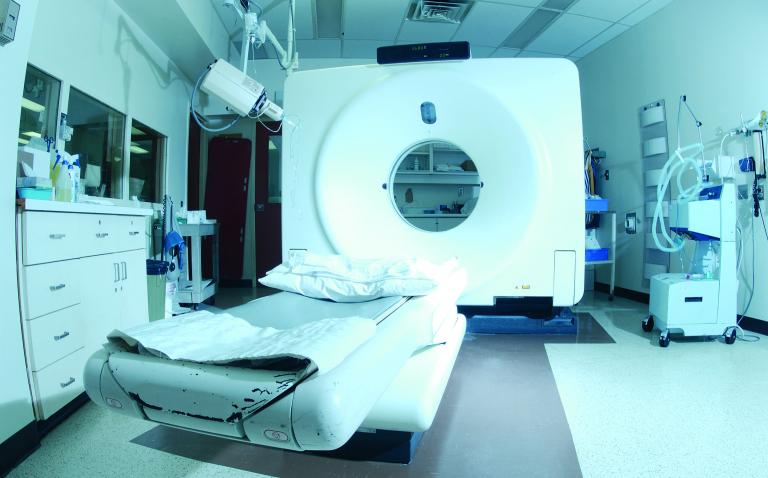
The “world’s first” dynamic volume CT (computerised tomography) scanner, which can scan a heart in a single heartbeat while administering just a fifth of the radiation dose of conventional scanners, was launched at the European Congress of Radiology (Vienna) yesterday.
Called the Aquilion ONE™, the new 320-slice CT machine is the first to allow radiologists to view continuous 4D (like video) real-time images of the heart and brain without the patient having to move up and down through the scanner.
With its 16cm detector – five times the size of traditional 64-slice CT scanners – and its dynamic volume CT imaging, clinicians will now be able to observe blood flow (perfusion), movement, and other functions of entire organs, and in precise detail. This coverage of the body will eliminate the
need to stitch together separate scans of organs that fit within the detector area.
Using the Aquilion ONE, cardiac scanning administers approximately 20% of the radiation dose of a 64-slice conventional CT scanner, and reduces radiation doses by 50% in scans for acute stroke.
“Until now, concerns regarding radiation have prevented doctors using more CT to assess the heart and brain,” says Dr Russell Bull, Consultant Radiologist at the Royal Bournemouth Hospital. “However, this technology allows very fast and accurate scanning of these organs using much lower radiation doses.”
The new technology will also offer additional benefits for doctors, patients, and hospital managers alike. The Aquilion ONE’s high resolution, dynamic volume imaging may reduce the need for multiple scans and invasive procedures, which are frequently more costly, time-consuming and less comfortable for the patient.
The Aquilion ONE scans extremely quickly, taking just one revolution (0.35 seconds) to scan an entire organ, unlike conventional systems, which revolve many times. For patients presenting with chest pain, the entire heart can be scanned in a single heartbeat.
The Aquilion ONE can also scan one area continuously, providing extremely quick and more precise information on the functionality of an organ. For patients presenting with acute stroke symptoms, just one examination taking no more than 60 seconds can provide critical information on blood flow through the brain including vascular analysis measures. This information could to be key to improving the rapid assessment and treatment of acute stroke.
Other organs can also be seen functioning, such as the lungs as the patient breathes in and out, and joints as the patient flexes them.
“With the volume scanning on the Aquilion ONE, I can see things that I never saw on any 64-slice scanner. This gives me unique diagnostic information,” says Dr Patrik Rogalla, Chief Radiologist and Director of the CT Division the Charité University Hospital, Berlin (Germany), the first centre to install the new scanner in Europe.
The Aquilion ONE took ten years to develop and at a cost of $500 million. The new system has recently been installed in an additional two European university hospital centres.


