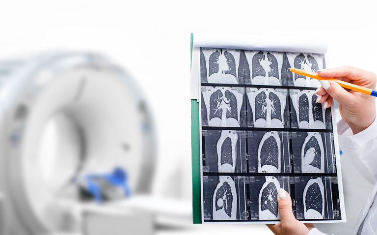
CT chest changes are less severe with the omicron COVID-19 variant compared to delta and help explain the better patient outcomes with delta
Computed tomography (CT) chest scan changes after infection with the omicron COVID-19 variant are less severe than those seen with the delta variant and lead to better patient outcomes. This was the conclusion of an analysis of CT chest scans by researchers from Oxford University, UK.
The COVID-19 omicron variant of concern was first identified in South Africa in November 2021 and an analysis in January 2022 suggested a significantly reduced chance of being hospitalised for individuals infected with the omicron variant compared to previous variants of concern. In fact, the authors suggested that this reduction in severity was most likely due due to a degree of protection from prior immunity.
While there is evidence to show that among those who experience a breakthrough COVID-19 infection, examination of CT chest scans indicate a lower level of pneumonia, the study did not provide any information on the infective variants involved. Moreover, a CT chest has become a valuable tool for the detection of both alternative diagnoses and complications of COVID-19, but to date, there is a lack of information on the CT changes induced after infection with different COVID-19 variants.
For the present study, the Oxford team compared the radiological patterns, imaging characteristics and disease severity on initial CT chest angiograms of patients infected with the omicron and delta variants of concern.
They retrospectively collected data on patients hospitalised and with a PCR confirmed positive COVID-19 test, using the S gene target failure as a surrogate for omicron infections. For the CT chest scans, a severity score (CT-SS) was calculated (ranging from 0 to 25, with higher scores reflecting greater severity) and CT imaging features such as bronchial wall thickening were also assessed.
Additional data collected included demographics, co-morbidities, laboratory findings, in-hospital treatments and clinical outcomes. Logistic regression models were created and adjusted for potential confounding variables to identify any differences in outcomes for the two variants.
CT chest changes induced by omicron and delta variants
A total of 106 adult patients with a mean age of 58 years (55% male) were included in the final analysis, of whom, 66 were infected with the delta variant.
Based on the CT chest findings, a higher proportion of scans were categorised as normal in patients with omicron compared to delta (37% vs 15%, omicron vs delta, p = 0.016). Interestingly, only 40% of those infected with omicron compared to 83% of those with delta, displayed the typical signs of pneumonia.
Patients infected with omicron had a lower median CT-SS score compared to those with delta (3.5 vs 11.8). Furthermore, bronchial wall thickening was more common in those with omicron (odds ratio, OR = 2.4, 95% CI 1.01 – 5.92, p = 0.04).
Infection with the delta variant was also associated with a higher odds of more severe infection (OR = 4.6, 95% CI 1.2 – 26, p = 0.01) and critical care admission (OR = 7).
The authors concluded that infection with the omicron variant is associated with fewer and less severe changes on a CT chest compared to the delta variant and led to better patient outcomes.
Citation
Tsakok MT et al. Chest CT and Hospital Outcomes in Patients with Omicron Compared with Delta Variant SARS-CoV-2 Infection Radiology 2022


