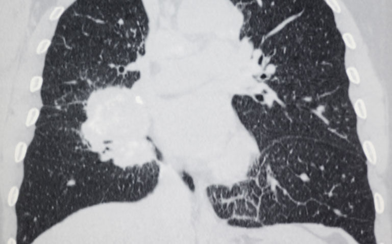
The presence of visual or quantitative emphysema on a CT-scan is associated with a more than twofold increased risk of developing lung cancer, according to a recent study
The detection of emphysema via visual or quantitative assessment on a CT-scan has been found to be linked with a higher odds of developing lung cancer.
This was the conclusion of a systematic review by researchers from the Departments of Epidemiology, Radiology and Pulmonary Diseases, University Medical Center Groningen, University of Groningen, Groningen, the Netherlands.
A chest computer tomography (CT) scan enables quantification of the amount of emphysema present in the lungs and while some evidence suggests that emphysema on a CT scan is related to lung cancer in a high-risk population, other data indicates that no CT measures of emphysema have an independent association with lung cancer.
With some uncertainty over the association between the presence of emphysema seen on a CT scan and lung cancer, for the present study, the researchers decided to undertake a systematic review and meta-analysis to further probe this association.
They searched all the major databases and included studies that specifically assessed the association between emphysema and the diagnosis of lung cancer based on histopathologic examination.
The team defined visual emphysema as disrupted lung vasculature and parenchyma with low attenuation occupying any lung zone on the chest CT scan and quantitative emphysema as the percentage of total lung volume below a given Hounsfield unit threshold (-950 HU at full inspiration). They also sought to examine whether the severity of emphysema was associated with lung cancer and graded this as trace, mild, moderate/severe.
The studies were stratified based on whether visual or quantitative assessments were used and the presence of confirmed lung cancer was the main outcome of interest expressed and expressed as an odds ratio, adjusted for age, gender and smoking status.
Emphysema and lung cancer risk
A total of 21 studies met the inclusion criteria with 3907 patients who had lung cancer and 103,175 controls.
The pooled odds ratio (OR) for lung cancer in the presence of emphysema was 2.3 (95% CI 2 – 2.6) in studies which employed visual assessment and 2.2 (95% CI 1.8 – 2.8) where the authors used quantitative assessment.
When stratified by disease severity, the overall pooled OR for lung cancer increased with disease severity although there were differences based on whether the data was acquired by visual or quantitative assessment.
For example, in studies that employed visual assessment, the ORs for lung cancer were 2.5 (trace disease), 3.7 (mild disease) and 4.5 (moderate to severe disease).
While these odds ratios were still elevated based on quantitative assessments, the magnitudes were slightly lower e.g., 1.9 for trace disease and 2.5 (moderate to severe disease).
Based on their findings, the author concluded that the presence of emphysema diagnosed on a chest CT scan was independently associated with a higher odds of developing lung cancer.
Citation
Yang X et al. Association between Chest CT–defined Emphysema and Lung Cancer: A Systematic Review and Meta-Analysis Radiology 2022


