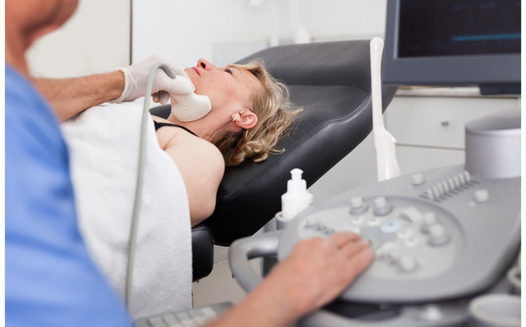
Ultrasonography is more accurate than clinical examination for LN metastases in squamous cell carcinoma (SCC) but has more false positives
Ultrasonography (US) is better than clinical examination for the detection of lymph node (LN) metastases in patients with squamous cell carcinoma (SCC) but is associated with higher rate of false positives. This was the conclusion of a study by a team from the Department of Dermatology, Erasmus Medical Center Cancer Institute, Rotterdam, the Netherlands.
Squamous cell carcinoma represents a malignant proliferation of the cutaneous epithelium and accounts for 20% to 50% of skin cancers. Fortunately however, the incidence of metastases with the cancer is low, with one UK study observing that after 36 months, 1.1% of cases in women and 2.5% in men become metastatic. Although a 2020 interdisciplinary European consensus guideline noted that imaging methods, for example, US are more sensitive than clinical examination, it added that ‘there are limited data on the use of US for nodal metastasis‘ for SCC.
Where clinical examination reveals suspicious findings, clinicians may request US although the added value of this imaging modality remains uncertain. For the present analysis, the Dutch researchers therefore examined the diagnostic accuracy of clinical examination and baseline US for the detection of LN metastases in patients with high risk cutaneous SCC of the head and neck. They also explored the accuracy of baseline US where baseline clinical examination did not suspect metastases.
The team retrospectively reviewed all consecutive, high risk patients at their centre. Where lymphadenopathy was detected upon examination or with one or more suspicious lymph nodes detected on US, a biopsy was undertaken and nodal metastasis was confirmed by cytology. The diagnostic accuracy of clinical examination and US was calculated based on sensitivity, specificity and positive predictive value (PPV).
Findings
During a 3-year period, 233 patients with a median age of 79.1 years (75.5% male) with 246 high-risk SCCs were included in the analysis.
Among the 246 tumours, 20 (8.1%) had suspicious lymphadenopathy upon clinical examination and 11 confirmed as LN metastatic after cytology. Suspicious lymph nodes were found on US in 28.5% (70/246) of cases and in 20 of these, metastasis was confirmed by cytology.
Using these data, the authors calculated the sensitivity of clinical examination to be 50% (95% CI 28 – 72%), with a specificity of 96% (95% CI 93 – 98%) and a PPV of 55%. In contrast, US had a sensitivity of 91% (95% CI 71 – 99%), a specificity of 78% (95% CI 72 – 83%) and a PPV of 29%.
In patients with a negative result upon clinical examination, 9 of 11 metastases were detected by US with a sensitivity of 82% although there were 45 false positives, making the PPV only 17%. Based on these findings, the authors suggested that while US examination was very sensitive, this needs to be seen in the context of a higher number of false positives.
The concluded that while US was a more sensitive screening tool to detect LN metastasis, the high false positive rate would in practice lead to a higher rate of unnecessary US and cytology procedures. They suggested that future studies should focus on identifying specific populations of those with SCCs who would benefit the most from baseline US to detect metastatic disease.
Citation
Tokez S et al. Assessment of the Diagnostic Accuracy of Baseline Clinical Examination and Ultrasonographic Imaging for the Detection of Lymph Node Metastasis in Patients With High-Risk Cutaneous Squamous Cell Carcinoma of the Head and Neck JAMA Dermatol 2021.


