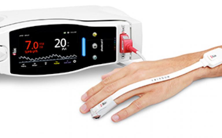
Masimo has announced the findings of a recently published study in which researchers at Bülent Ecevit University in Zonguldak, Turkey compared two noninvasive methods of predicting fluid responsiveness in mechanically ventilated patients in the intensive care unit (ICU): Masimo PVi® (pleth variability index, measured non-invasively and continuously using SET® pulse oximetry sensors) and dIVC (distensibility index of inferior vena cava, measured non-invasively by radiologists using an ultrasound machine and probe).1
In the study, Drs. Pişkin and Öz sought to compare the performance of the two methods by enrolling 72 mechanically ventilated patients and taking various measurements before and after passive leg raising (PLR). In addition to PVi (measured with Masimo Radical-7® Pulse CO-Oximeters®) and dIVC (measured by a radiologist using Esaote MyLab 30), central venous pressure (CVP) and cardiac index (CI) were measured. Patients who exhibited a greater than 15% increase in CI attributable to the PLR manoeuver were classified as volume responders, and those with less than 15% or no change, as non-responders.

The researchers found that Masimo noninvasive and continuous PVi, at a threshold value of >14%, provided 95% sensitivity and 81.2% specificity (p<0.001, AUC = 0.939 (0.857-0.982)), which was statistically significant. Ultrasound, non-invasive dIVC, at a threshold value of >23.8%, provided 80% sensitivity and 87.5% specificity (p<0.001, AUC = 0.928 (0.842-0.975)), which was also statistically significant. The invasive method, CVP, at a threshold value of ≤7 mmHg, provided 70% sensitivity and 53.1% specificity to predict fluid responsiveness (p=0.066, Area Under the Curve = 0.622 (0.500-0.724)), which was not statistically significant.
The investigators noted that “our results show that non-invasively assessed PVi and dIVC were good predictors of fluid responsiveness after PLR in ICU patients on mechanical ventilation. By contrast, the invasively assessed CVP was a poor predictor of fluid responsiveness as a static variable of cardiac preload.” They concluded that, “Both PVi and dIVC may be used to identify the fluid responsiveness of all ICU patients undergoing continuous treatment linked to mechanical ventilation; both methods are easily applied, non-invasive, and can be performed at the bedside.”
Reference
- Pişkin Ö and Öz I. Accuracy of pleth variability index compared with inferior vena cava diameter to predict fluid responsiveness in mechanically ventilated patients. Medicine. (2017) 96:47(e8889).


