


Point-of-care ultrasound (POCUS) has become an important adjunct to clinical diagnosis and procedural guidance in the Pediatric Emergency Department (PED), supported by literature demonstrating that its use can improve patient safety and expedite lifesaving care. POCUS further helps to reduce costs and children’s exposure to ionizing radiation. Not only is POCUS ideally suited for “rule-in” diagnostic applications for many pediatric indications, but ultrasound guidance can also improve the safety and success of common procedures, including placement of central venous catheters (CVCs) and peripheral intravenous (PIV) catheters, thoracentesis, paracentesis, regional anesthesia, lumbar punctures and other procedures.[1]
Moreover, POCUS can aid in the evaluation of musculoskeletal pathology including fractures, soft tissue injuries and joint effusions, and help guide arthrocentesis.[2] This review provides an overview of a few of the important diagnostic and procedural applications of POCUS in Pediatric Emergency Medicine (PEM) with case examples.
Using Ultrasound-Guided Vascular Access to Achieve the “One-Stick Standard”
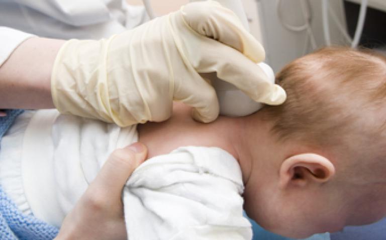
One of the most common procedures performed in medicine, overall, is vascular access; its success can be lifesaving for many children who present to the ED. The Agency for Healthcare Research and Quality has identified “use of real-time ultrasound guidance during central line insertion to prevent complications” as one of the 12 most highly rated patient safety practices.[3]
Ultrasound-guided CVC, rather than the traditional blind landmark approach has become the standard of care. In a 2016 policy statement, the American College of Emergency Physicians (ACEP) encourages a new goal for vascular access: the “one-stick standard.” To achieve it, ACEP recommends the use of POCUS for both CVC and PIV catheter placement, listing such advantages as “improved patient safety, decreased procedural attempts and decreased time to perform many procedures in patients [for] whom the technique would otherwise be difficult.”
Pediatric patients, particularly those who are smaller, dehydrated or chronically ill, can present unique challenges with successfully obtaining vascular access. Ultrasound-guided PIV insertions can help difficult-access patients avoid delays in care and reduce the need for CVCs, which can have serious risks, including pneumothorax.[4] In a randomized controlled trial comparing ultrasound-guided PIV placement with traditional methods in difficult-access pediatric patients, our team demonstrated that ultrasound guidance had a superior success rate (80% vs. 64%) and was faster (6.3 vs. 14.4 minutes), with fewer attempts (median: 1 vs. 3), and fewer needle redirections (median: 2 vs. 10).[5]
An Accurate, Cost-Effective Initial Test for Suspected Pediatric Appendicitis
Acute appendicitis is the most common surgical emergency in children. The American College of Radiology’s Appropriateness Criteria recommends ultrasound as the “preferred” and “most cost-effective” initial test for suspected pediatric appendicitis.6 The use of radiology-performed ultrasound as the first diagnostic test in a staged imaging algorithm, has been shown to significantly reduce pediatric exposure to ionizing radiation without diminishing diagnostic accuracy. [7,8]
Our team recently conducted the first study of a staged diagnostic algorithm for pediatric appendicitis that included POCUS, using a convenience sampling of patients 2 to 18 years of age who presented to our pediatric ED.[9] An algorithm was followed: POCUS was the first study performed, followed by radiology-performed ultrasound. If the ultrasound results were equivocal, the patient then also received a CT scan. We demonstrated a sensitivity of 93.8% and a specificity of 87.5% for POCUS versus 81.25% and 100% respectively for radiology-performed ultrasound. Most compelling was the finding that with this staged algorithm, the use of CT scans was decreased by 55%. In addition, POCUS can also identify other bowel pathologies.
We recently presented two cases in which pediatric patients examined for suspected appendicitis were found to have multiple ingested magnets, resulting in a subsequent bowel perforation.[10] The foreign bodies, which were visualized with POCUS and radiographs, were successfully removed and the intestinal perforations repaired in the operating room, with good outcomes for both patients. Small children are particularly at high risk for occult foreign body ingestions, and these cases highlight the potential of ultrasound to reveal an unexpected surgical emergency
Nearly Perfect Accuracy for Diagnosing Intussusception
In children 6 months to 2 years of age, intussusception is the leading surgical cause of abdominal obstruction [11], and can lead to serious or fatal complications if the diagnosis and treatment are delayed. Due to its ease of performance, safety and reported sensitivities of 98-100% and specificities of 88-100% when performed by radiologists [12, 13, 14], ultrasound has become the initial diagnostic test of choice. Recently, POCUS has been used for this purpose as well, with a recent study revealing that point-of-care ultrasound has a sensitivity of 85% and a specificity of 97% for the evaluation of suspected intussusception.[15] Ultrasonography can also be used to identify features that might suggest whether the enema reduction will be successful or not. Some of these sonographic features include the absence of blood –flow to the affected bowel, or the presence of surrounding free fluid.[16]
Sparing Children With Soft-Tissue Infections Needless Incision & Drainage and Sedation Emergency
Department visits for skin and soft-tissue infections have tripled since the 1990s.[17] However, the clinical history and physical examination alone are often unreliable in distinguishing cellulitis from abscess. The correct diagnosis is crucial, since treating an abscess requires incision and drainage, a procedure that often necessitates sedation in pediatric patients. Conversely, if an abscess goes undetected, the result can be a more severe infection and return ED visits for treatment.
In multiple prospective pediatric studies, ultrasound visualization was superior to physical examination alone for the accurate diagnosis of soft-tissue infections. 18,19 Sivitz et al. reported that POCUS changed management in 22% of cases, either by identifying abscesses that needed treatment, or by sparing children with superficial infections unnecessary invasive procedures and sedation. For detecting ‑fluid requiring incision and drainage, ultrasound had a sensitivity of 90% and a specfiicity of 83%, versus 75% and 80% respectively for the physical examination alone.
Comparable or Superior Accuracy to X-ray for Detecting Foreign Bodies
POCUS has an important role in evaluating children with suspected soft-tissue foreign bodies—a common scenario in the PED. Friedman et al. studied 131 wounds in patients with suspected foreign bodies and found no statistically signicant difference in the diagnostic accuracy of radiographs when compared with ultrasound alone.[20] However, ultrasound is superior to radiographs for detection of radiolucent foreign body materials, such as plastic and wood, as opposed to radiopaque materials (metal and glass).
A colleague and I published a case report demonstrating how easily and effectively ultrasound can be utilized for such evaluation. A wooden splinter in a 10-year-old boy’s finger that would have been invisible on radiograph, was clearly visualized under POCUS, which also helped guide removal of the foreign body.[21]
A Promising Alternative to X-rays for Rapid Diagnosis of Pneumonia
Pneumonia is the single largest infectious cause of childhood mortality worldwide.[22] The use of lung ultrasound (LUS) to diagnose childhood pneumonia is an important, emerging application, particularly in resource-limited settings.
A recent meta-analysis revealed that LUS has an overall pooled sensitivity of 96% and specificity of 93% for diagnosing pneumonia, leading the investigators to conclude that, “Current evidence supports LUS as an imaging alternative for the diagnosis of childhood pneumonia.”[23] In addition, LUS enables the rapid diagnosis of other pulmonary pathologies, facilitating the care of sick children. In a study of children and young adults evaluated for suspected pneumonia, Shah et al. reported a mean ultrasonography examination time of 7 minutes, with a 97% specificity for lung consolidations exceeding 1 cm.
For the evaluation of complex pleural effusions or empyemas in children, chest CT can be replaced with a POCUS, with or without a chest radiograph.[24]
Conclusions
Research into novel applications of POCUS in PEM is rapidly expanding, fueled by the growing recognition that pediatric patients are not simply small adults. It is an exciting time in medicine, as the unique needs of the youngest, most vulnerable patients have inspired an astonishing variety of innovations and discoveries aimed at accelerating the diagnoses of life-threatening conditions and guiding safer, more successful care. With point-of-care ultrasound and the crucial information it provides, PEM physicians are ideally equipped to help critically ill or injured children achieve optimal outcomes.
Author bio
Stephanie J Doniger, MD, RDMS, FAAP, FACEP is one of the leading experts in pediatric point-of-care ultrasound. Following her training in Pediatrics and Pediatric Emergency Medicine, she was the rst to complete an additional Emergency Ultrasound Fellowship in 2008. Since that time, Dr. Doniger has authored and co-authored numerous book chapters, articles and other publications on point-of-care ultrasound. She edited the rst textbook “Pediatric Emergency and Critical Care Ultrasound,” and has lectured on national and international levels in both English and Spanish. Dr. Doniger has held multiple leadership positions, including the Subcommittee Chair of the ACEP Pediatric Ultrasound Subcommittee for ve years and the WINFOCUS Board of Directors. She has been a member of WINFOCUS since 2008 and is currently in charge of organizing pediatric point-of-care ultrasound education throughout Latin America.
References
1. Moore CL, Copel JA. Point-of-care ultrasonography. N Engl J Med. 2011; 64(8):749–757.
2. Marin JR, Abo AM, Arroyo AC et al. Pediatric emergency medicine point-of-care ultrasound: summary of the evidence. Crit Ultrasound J (2016) 8:16.
3. Making health care safer: a critical analysis of patient safety practices. Rockville, MD: Agency for Healthcare Research and Quality. (AHRQ publication no. 01-E058.)
4. Chiang V, Baskin M. Uses and complications of central venous catheters inserted in a pediatric emergency department. Pediatr Emerg Care. 2000;16:230Y232.
5. Doniger SJ, Ishimine P et al. Randomized Controlled Trial of Ultrasound-Guided Peripheral Intravenous Catheter Placement Versus Traditional Techniques in Difcult-Access Pediatric Patients. Pediatr Emerg Care. 2009 Mar;25(3):154-9.
6. American College of Radiology website. ACR Appropriateness Criteria Right Lower Quadrant Pain—Suspected Appendicitis. Accessed on January 15, 2018 at https://acsearch.acr.org/docs/69357/Narrative/.
7. Krishnamoorthi R, Ramarajan N, Wang NE, et al. Effectiveness of a staged US and CT protocol for the diagnosis of pediatric appendicitis: reducing radiation exposure in the age of ALARA. Radiology. 2011;259: 231–239.
8. Ramarajan N, Krishnamoorthi R, Barth R, et al. An interdisciplinary initiative to reduce radiation exposure: evaluation of appendicitis in a pediatric emergency department with clinical assessment supported by a staged ultrasound and computed tomography pathway. Acad Emerg Med. 2009;16:1258–1265.
9. Doniger SJ and Kornblith A. Point-of-Care Ultrasound Integrated Into a Staged Diagnostic Algorithm for Pediatric Appendicitis. Pediatr Emerg Care. 2016; Jun 14 9 [Epub ahead of print].
10. Leibowvich S, Doniger SJ. The Use of Point-of-Care Ultrasound to Evaluate for Intestinal Foreign Bodies in the Pediatric Emergency Department. Pediatr Emerg Care. 2013 Jul;29(7):870-3.
11. Wyllie R. Ileus, adhesions, intussusception, and closed-loop obstructions. In: Kliegman R, Nelson WE, eds. Nelson Textbook of Pediatrics. 18th ed. Philadelphia, PA: Saunders; 2007:lii, 3147.
12. Verschelden P, Filiatrault D, Garel L, Grignon A, Perreault G, Boisvert J, Dubois J (1992) Intussusception in children: reliability of US in diagnosis—a prospective study. Radiology 184:741–744
13. Daneman A, Navarro O (2004) Intussusception. Part 2: an update on the evolution of management. Pediatr Radiol 34:97–108 (quiz 187)
14. Navarro O, Daneman A (2004) Intussusception. Part 3: diagnosis and management of those with an identiable or predisposing cause and those that reduce spontaneously. Pediatr Radiol 34:305–312 (quiz 369)
15. Riera A, Hsiao AL, Langhan ML et al. Diagnosis of intussusception by physician novice sonographers in the emergency department. Ann Emerg Med. 2012; 60(3):264–268.
16. Marin JF, Abo AM, Orroyo AC, Doniger SJ et al. Pediatric emergency medicine point-of-care ultrasound: summary of the evidence. Crit Ultrasound J. (2016) 8:16.
17. Pallin DJ, Egan DJ, Pelletier AJ, Espinola JA, Hooper DC, Camargo CA Jr. Increased US emergency department visits for skin and soft tissue infections, and changes in antibiotic choices, during the emergence of community-associated methicillin-resistant Staphylococcus aureus. Ann Emerg Med. 2008;51(3):291-298.
18. Marin JR, Bilker W, Lautenbach E, Alpern ER. Reliability of clinical examinations for pediatric skin and soft-tissue infections. Pediatrics. 2010;126(5): 925-930.
19. Sivitz AB, Lam SH, Ramirez-Schrempp D, Valente JH, Nagdev AD. Effect of bedside ultrasound on management of pediatric soft-tissue infection. J Emerg Med. 2010;39(5):637-643.
20. Friedman DI, Forti RJ, Wall SP, Crain EF. The utility of bedside ultrasound and patient perception in detecting soft tissue foreign bodies in children. Pediatr Emerg Care 2005;21(8):487–492.
21. Teng M, Doniger SJ. Subungual wooden splinter visualized with bedside sonography. Pediatr Emerg Care. 2012;28(4):392–394.
22. World Health Organization. Pneumonia. Fact Sheet No 331. Geneva, Switzerland. Available from: www.who.int/mediacentre/factsheets/fs331/en/. Accessed January 12, 2018.
23. Pereda MA, Chavez MA, Hooper-Miele CC, et al. Lung ultrasound for the diagnosis of pneumonia in children: a meta-analysis. Pediatrics. 2015;135(4):714–722.
24. Hajalioghli P, Nemati M, Dinprast Saleh L, Fouladi DF. Can chest computed tomography be replaced by lung ultrasonography with or without plain chest radiography in pediatric pneumonia? J Thorac Imaging. 2016;31(4):247–252.
